Abdullah S Alqahtani, Wasan M Alshareef, Hanan T Aljadani, Wesal O Hawsawi, Marya H Shaheen
{"title":"人工智能在糖尿病视网膜病变筛查中的疗效:系统回顾和荟萃分析。","authors":"Abdullah S Alqahtani, Wasan M Alshareef, Hanan T Aljadani, Wesal O Hawsawi, Marya H Shaheen","doi":"10.1186/s40942-025-00670-9","DOIUrl":null,"url":null,"abstract":"<p><strong>Background: </strong>To evaluate the efficacy of artificial intelligence (AI) in screening for diabetic retinopathy (DR) using fundus images and optical coherence tomography (OCT) in comparison to traditional screening methods.</p><p><strong>Methods: </strong>This systematic review was registered with PROSPERO (ID: CRD42024560750). Systematic searches were conducted in PubMed Medline, Cochrane Central, ScienceDirect, and Web of Science using keywords such as \"diabetic retinopathy,\" \"screening,\" and \"artificial intelligence.\" Only studies published in English from 2019 to July 22, 2024, were considered. We also manually reviewed the reference lists of relevant reviews. Two independent reviewers assessed the risk of bias using the QUADAS-2 tool, resolving disagreements through discussion with the principal investigator. Meta-analysis was performed using MetaDiSc software (version 1.4). To calculate combined sensitivity, specificity, summary receiver operating characteristic (SROC) plots, forest plots, and subgroup analyses were performed according to clinician type (ophthalmologists vs. retina specialists) and imaging modality (fundus images vs. fundus images + OCT).</p><p><strong>Results: </strong>18 studies were included. Meta-analysis showed that AI systems demonstrated superior diagnostic performance compared to doctors, with the pooled sensitivity, specificity, diagnostic odds ratio, and Cochrane Q index of the AI being 0.877, 0.906, 0.94, and 153.79 accordingly. The Fagan nomogram analysis further confirmed the strong diagnostic value of AI. Subgroup analyses revealed that factors like imaging modality, and doctor expertise can influence diagnostic performance.</p><p><strong>Conclusion: </strong>AI systems have demonstrated strong diagnostic performance in detecting diabetic retinopathy, with sensitivity and specificity comparable to or exceeding traditional clinicians.</p>","PeriodicalId":14289,"journal":{"name":"International Journal of Retina and Vitreous","volume":"11 1","pages":"48"},"PeriodicalIF":2.4000,"publicationDate":"2025-04-22","publicationTypes":"Journal Article","fieldsOfStudy":null,"isOpenAccess":false,"openAccessPdf":"https://www.ncbi.nlm.nih.gov/pmc/articles/PMC12012971/pdf/","citationCount":"0","resultStr":"{\"title\":\"The efficacy of artificial intelligence in diabetic retinopathy screening: a systematic review and meta-analysis.\",\"authors\":\"Abdullah S Alqahtani, Wasan M Alshareef, Hanan T Aljadani, Wesal O Hawsawi, Marya H Shaheen\",\"doi\":\"10.1186/s40942-025-00670-9\",\"DOIUrl\":null,\"url\":null,\"abstract\":\"<p><strong>Background: </strong>To evaluate the efficacy of artificial intelligence (AI) in screening for diabetic retinopathy (DR) using fundus images and optical coherence tomography (OCT) in comparison to traditional screening methods.</p><p><strong>Methods: </strong>This systematic review was registered with PROSPERO (ID: CRD42024560750). Systematic searches were conducted in PubMed Medline, Cochrane Central, ScienceDirect, and Web of Science using keywords such as \\\"diabetic retinopathy,\\\" \\\"screening,\\\" and \\\"artificial intelligence.\\\" Only studies published in English from 2019 to July 22, 2024, were considered. We also manually reviewed the reference lists of relevant reviews. Two independent reviewers assessed the risk of bias using the QUADAS-2 tool, resolving disagreements through discussion with the principal investigator. Meta-analysis was performed using MetaDiSc software (version 1.4). To calculate combined sensitivity, specificity, summary receiver operating characteristic (SROC) plots, forest plots, and subgroup analyses were performed according to clinician type (ophthalmologists vs. retina specialists) and imaging modality (fundus images vs. fundus images + OCT).</p><p><strong>Results: </strong>18 studies were included. Meta-analysis showed that AI systems demonstrated superior diagnostic performance compared to doctors, with the pooled sensitivity, specificity, diagnostic odds ratio, and Cochrane Q index of the AI being 0.877, 0.906, 0.94, and 153.79 accordingly. The Fagan nomogram analysis further confirmed the strong diagnostic value of AI. Subgroup analyses revealed that factors like imaging modality, and doctor expertise can influence diagnostic performance.</p><p><strong>Conclusion: </strong>AI systems have demonstrated strong diagnostic performance in detecting diabetic retinopathy, with sensitivity and specificity comparable to or exceeding traditional clinicians.</p>\",\"PeriodicalId\":14289,\"journal\":{\"name\":\"International Journal of Retina and Vitreous\",\"volume\":\"11 1\",\"pages\":\"48\"},\"PeriodicalIF\":2.4000,\"publicationDate\":\"2025-04-22\",\"publicationTypes\":\"Journal Article\",\"fieldsOfStudy\":null,\"isOpenAccess\":false,\"openAccessPdf\":\"https://www.ncbi.nlm.nih.gov/pmc/articles/PMC12012971/pdf/\",\"citationCount\":\"0\",\"resultStr\":null,\"platform\":\"Semanticscholar\",\"paperid\":null,\"PeriodicalName\":\"International Journal of Retina and Vitreous\",\"FirstCategoryId\":\"1085\",\"ListUrlMain\":\"https://doi.org/10.1186/s40942-025-00670-9\",\"RegionNum\":0,\"RegionCategory\":null,\"ArticlePicture\":[],\"TitleCN\":null,\"AbstractTextCN\":null,\"PMCID\":null,\"EPubDate\":\"\",\"PubModel\":\"\",\"JCR\":\"Q2\",\"JCRName\":\"OPHTHALMOLOGY\",\"Score\":null,\"Total\":0}","platform":"Semanticscholar","paperid":null,"PeriodicalName":"International Journal of Retina and Vitreous","FirstCategoryId":"1085","ListUrlMain":"https://doi.org/10.1186/s40942-025-00670-9","RegionNum":0,"RegionCategory":null,"ArticlePicture":[],"TitleCN":null,"AbstractTextCN":null,"PMCID":null,"EPubDate":"","PubModel":"","JCR":"Q2","JCRName":"OPHTHALMOLOGY","Score":null,"Total":0}
引用次数: 0
摘要
背景:评价人工智能(AI)在眼底图像和光学相干断层扫描(OCT)筛查糖尿病视网膜病变(DR)中的应用效果,并与传统筛查方法进行比较。方法:本系统评价在PROSPERO注册(ID: CRD42024560750)。在PubMed Medline、Cochrane Central、ScienceDirect和Web of Science等网站上进行了系统搜索,关键词包括“糖尿病视网膜病变”、“筛查”和“人工智能”。仅考虑2019年至2024年7月22日期间发表的英文研究。我们还手动审查了相关审查的参考列表。两名独立审稿人使用QUADAS-2工具评估偏倚风险,通过与主要研究者讨论解决分歧。meta分析采用MetaDiSc软件(版本1.4)进行。根据临床医生类型(眼科医生vs视网膜专家)和成像方式(眼底图像vs眼底图像+ OCT)进行综合敏感性、特异性、总受试者操作特征(SROC)图、森林图和亚组分析。结果:纳入18项研究。meta分析显示,人工智能系统的诊断性能优于医生,人工智能系统的综合敏感性、特异性、诊断优势比和Cochrane Q指数分别为0.877、0.906、0.94和153.79。Fagan模态图分析进一步证实了AI较强的诊断价值。亚组分析显示,成像方式和医生专业知识等因素可以影响诊断表现。结论:人工智能系统在检测糖尿病视网膜病变方面表现出强大的诊断能力,其敏感性和特异性与传统临床医生相当或超过传统临床医生。
The efficacy of artificial intelligence in diabetic retinopathy screening: a systematic review and meta-analysis.
Background: To evaluate the efficacy of artificial intelligence (AI) in screening for diabetic retinopathy (DR) using fundus images and optical coherence tomography (OCT) in comparison to traditional screening methods.
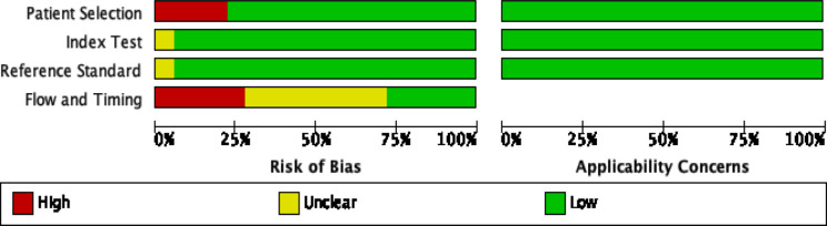
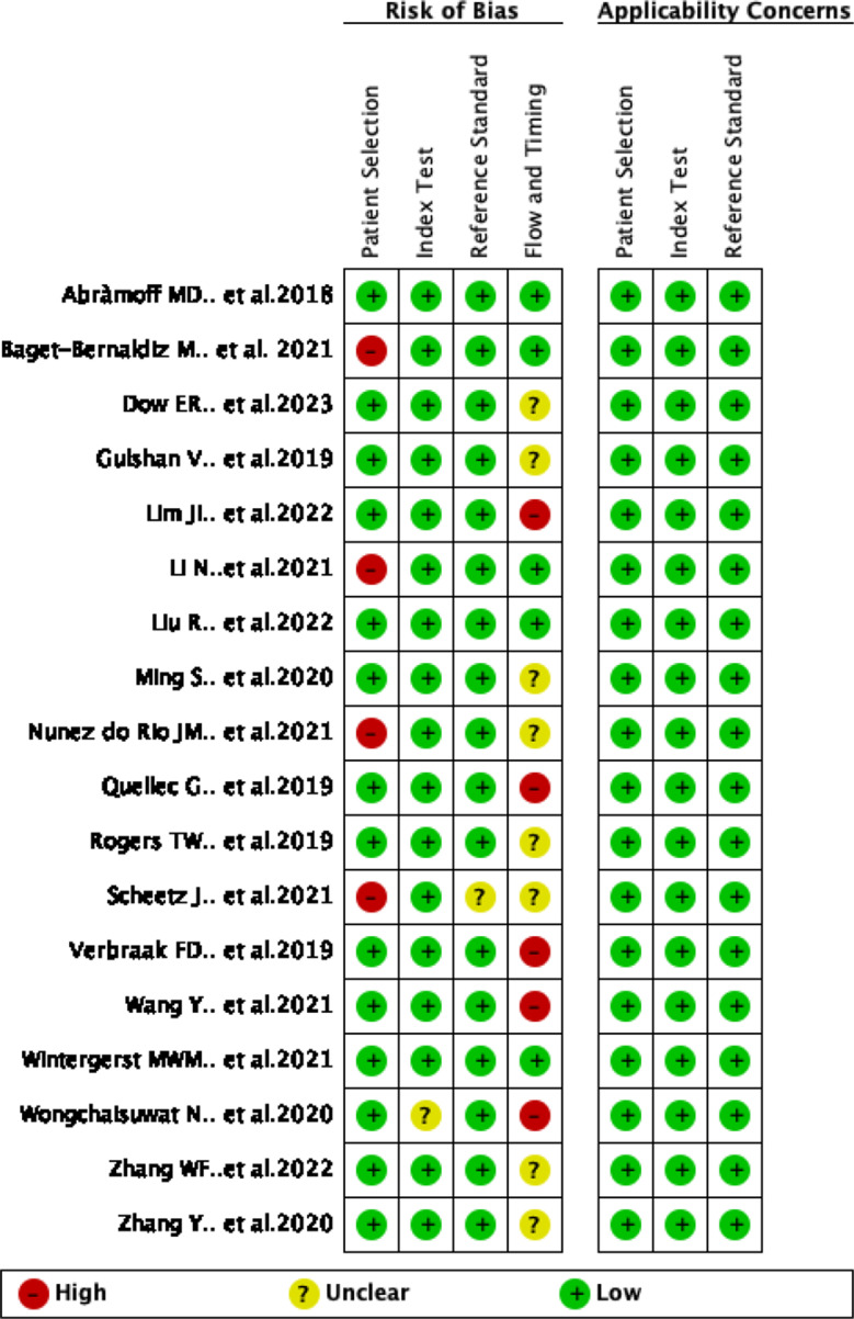
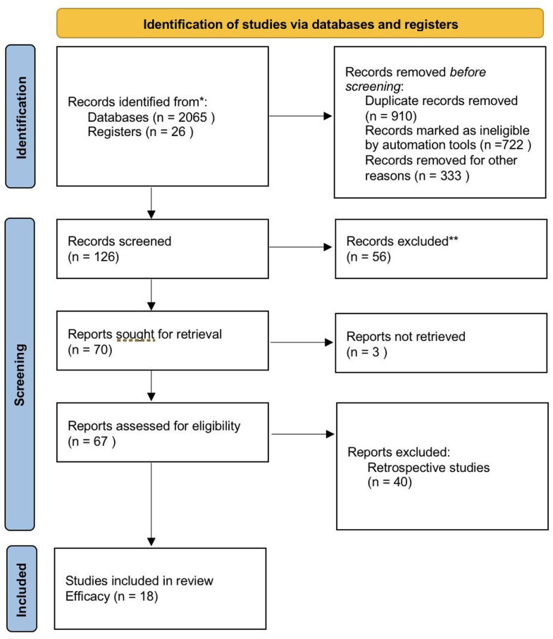
Methods: This systematic review was registered with PROSPERO (ID: CRD42024560750). Systematic searches were conducted in PubMed Medline, Cochrane Central, ScienceDirect, and Web of Science using keywords such as "diabetic retinopathy," "screening," and "artificial intelligence." Only studies published in English from 2019 to July 22, 2024, were considered. We also manually reviewed the reference lists of relevant reviews. Two independent reviewers assessed the risk of bias using the QUADAS-2 tool, resolving disagreements through discussion with the principal investigator. Meta-analysis was performed using MetaDiSc software (version 1.4). To calculate combined sensitivity, specificity, summary receiver operating characteristic (SROC) plots, forest plots, and subgroup analyses were performed according to clinician type (ophthalmologists vs. retina specialists) and imaging modality (fundus images vs. fundus images + OCT).
Results: 18 studies were included. Meta-analysis showed that AI systems demonstrated superior diagnostic performance compared to doctors, with the pooled sensitivity, specificity, diagnostic odds ratio, and Cochrane Q index of the AI being 0.877, 0.906, 0.94, and 153.79 accordingly. The Fagan nomogram analysis further confirmed the strong diagnostic value of AI. Subgroup analyses revealed that factors like imaging modality, and doctor expertise can influence diagnostic performance.
Conclusion: AI systems have demonstrated strong diagnostic performance in detecting diabetic retinopathy, with sensitivity and specificity comparable to or exceeding traditional clinicians.
期刊介绍:
International Journal of Retina and Vitreous focuses on the ophthalmic subspecialty of vitreoretinal disorders. The journal presents original articles on new approaches to diagnosis, outcomes of clinical trials, innovations in pharmacological therapy and surgical techniques, as well as basic science advances that impact clinical practice. Topical areas include, but are not limited to: -Imaging of the retina, choroid and vitreous -Innovations in optical coherence tomography (OCT) -Small-gauge vitrectomy, retinal detachment, chromovitrectomy -Electroretinography (ERG), microperimetry, other functional tests -Intraocular tumors -Retinal pharmacotherapy & drug delivery -Diabetic retinopathy & other vascular diseases -Age-related macular degeneration (AMD) & other macular entities

 求助内容:
求助内容: 应助结果提醒方式:


