Yuen Keat Gan, Hong Kee Ng, William H. Morgan, Dao-Yi Yu
{"title":"交联明胶支架的长期形态学改变。","authors":"Yuen Keat Gan, Hong Kee Ng, William H. Morgan, Dao-Yi Yu","doi":"10.1111/ceo.14545","DOIUrl":null,"url":null,"abstract":"<div>\n \n \n <section>\n \n <h3> Background</h3>\n \n <p>This study investigates the long-term morphological outcomes of crosslinked gelatin stents over a 10-year period.</p>\n </section>\n \n <section>\n \n <h3> Methods</h3>\n \n <p>Participants from the FDA phase 1 and 2 trials for the XEN 140 stent were recalled for follow-up examinations between 9 and 12 years after their initial implantation. The stent area was evaluated using anterior segment optical coherence tomography (AS-OCT) in conjunction with clinical assessments. We focused on measuring specific alterations in stent dimensions, such as external surface pitting or ‘nibbling’, complete wall breaches and luminal obstruction.</p>\n </section>\n \n <section>\n \n <h3> Results</h3>\n \n <p>We evaluated 11 eyes from 9 patients, with an average follow-up period of 11.2 years (SD ± 0.7). The mean preoperative intraocular pressure (IOP) was 22.5 mmHg (SD ± 4.9), which decreased to 14.7 mmHg (SD ± 1.2) a decade after surgery. Notably, four eyes no longer required antiglaucoma medication. Out of the 11 cases, only 8 yielded image quality suitable for analysis, all of which exhibited stent wall nibbling. The average nibbling depth was 70.17 μm (SD ± 36) in the intrascleral area, 79.5 μm (SD ± 18) in the proximal conjunctival region and 42.25 μm (SD ± 5) in the distal conjunctival region. Nibbling was most frequent in the proximal conjunctival area with 26 instances (<i>p</i> = 0.0006), followed by the intrascleral area, 21, and the distal conjunctival section, 9. Hyperreflective lumens were noted in five cases, with one complete stent wall discontinuity.</p>\n </section>\n \n <section>\n \n <h3> Conclusion</h3>\n \n <p>In cases where imaging provided clear visibility, the stents exhibited signs of morphometric changes after 11 years. This degradation process seems to initiate externally, leading to luminal obstruction and eventual stent failure.</p>\n </section>\n </div>","PeriodicalId":55253,"journal":{"name":"Clinical and Experimental Ophthalmology","volume":"53 6","pages":"611-618"},"PeriodicalIF":5.6000,"publicationDate":"2025-04-28","publicationTypes":"Journal Article","fieldsOfStudy":null,"isOpenAccess":false,"openAccessPdf":"https://onlinelibrary.wiley.com/doi/epdf/10.1111/ceo.14545","citationCount":"0","resultStr":"{\"title\":\"Long-Term Morphological Changes of Crosslinked Gelatin Stent\",\"authors\":\"Yuen Keat Gan, Hong Kee Ng, William H. Morgan, Dao-Yi Yu\",\"doi\":\"10.1111/ceo.14545\",\"DOIUrl\":null,\"url\":null,\"abstract\":\"<div>\\n \\n \\n <section>\\n \\n <h3> Background</h3>\\n \\n <p>This study investigates the long-term morphological outcomes of crosslinked gelatin stents over a 10-year period.</p>\\n </section>\\n \\n <section>\\n \\n <h3> Methods</h3>\\n \\n <p>Participants from the FDA phase 1 and 2 trials for the XEN 140 stent were recalled for follow-up examinations between 9 and 12 years after their initial implantation. The stent area was evaluated using anterior segment optical coherence tomography (AS-OCT) in conjunction with clinical assessments. We focused on measuring specific alterations in stent dimensions, such as external surface pitting or ‘nibbling’, complete wall breaches and luminal obstruction.</p>\\n </section>\\n \\n <section>\\n \\n <h3> Results</h3>\\n \\n <p>We evaluated 11 eyes from 9 patients, with an average follow-up period of 11.2 years (SD ± 0.7). The mean preoperative intraocular pressure (IOP) was 22.5 mmHg (SD ± 4.9), which decreased to 14.7 mmHg (SD ± 1.2) a decade after surgery. Notably, four eyes no longer required antiglaucoma medication. Out of the 11 cases, only 8 yielded image quality suitable for analysis, all of which exhibited stent wall nibbling. The average nibbling depth was 70.17 μm (SD ± 36) in the intrascleral area, 79.5 μm (SD ± 18) in the proximal conjunctival region and 42.25 μm (SD ± 5) in the distal conjunctival region. Nibbling was most frequent in the proximal conjunctival area with 26 instances (<i>p</i> = 0.0006), followed by the intrascleral area, 21, and the distal conjunctival section, 9. Hyperreflective lumens were noted in five cases, with one complete stent wall discontinuity.</p>\\n </section>\\n \\n <section>\\n \\n <h3> Conclusion</h3>\\n \\n <p>In cases where imaging provided clear visibility, the stents exhibited signs of morphometric changes after 11 years. This degradation process seems to initiate externally, leading to luminal obstruction and eventual stent failure.</p>\\n </section>\\n </div>\",\"PeriodicalId\":55253,\"journal\":{\"name\":\"Clinical and Experimental Ophthalmology\",\"volume\":\"53 6\",\"pages\":\"611-618\"},\"PeriodicalIF\":5.6000,\"publicationDate\":\"2025-04-28\",\"publicationTypes\":\"Journal Article\",\"fieldsOfStudy\":null,\"isOpenAccess\":false,\"openAccessPdf\":\"https://onlinelibrary.wiley.com/doi/epdf/10.1111/ceo.14545\",\"citationCount\":\"0\",\"resultStr\":null,\"platform\":\"Semanticscholar\",\"paperid\":null,\"PeriodicalName\":\"Clinical and Experimental Ophthalmology\",\"FirstCategoryId\":\"3\",\"ListUrlMain\":\"https://onlinelibrary.wiley.com/doi/10.1111/ceo.14545\",\"RegionNum\":2,\"RegionCategory\":\"医学\",\"ArticlePicture\":[],\"TitleCN\":null,\"AbstractTextCN\":null,\"PMCID\":null,\"EPubDate\":\"\",\"PubModel\":\"\",\"JCR\":\"Q1\",\"JCRName\":\"OPHTHALMOLOGY\",\"Score\":null,\"Total\":0}","platform":"Semanticscholar","paperid":null,"PeriodicalName":"Clinical and Experimental Ophthalmology","FirstCategoryId":"3","ListUrlMain":"https://onlinelibrary.wiley.com/doi/10.1111/ceo.14545","RegionNum":2,"RegionCategory":"医学","ArticlePicture":[],"TitleCN":null,"AbstractTextCN":null,"PMCID":null,"EPubDate":"","PubModel":"","JCR":"Q1","JCRName":"OPHTHALMOLOGY","Score":null,"Total":0}
Long-Term Morphological Changes of Crosslinked Gelatin Stent
Background
This study investigates the long-term morphological outcomes of crosslinked gelatin stents over a 10-year period.
Methods
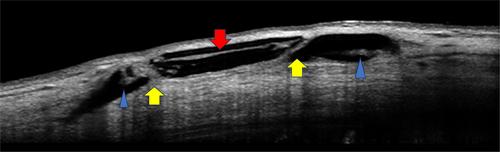
Participants from the FDA phase 1 and 2 trials for the XEN 140 stent were recalled for follow-up examinations between 9 and 12 years after their initial implantation. The stent area was evaluated using anterior segment optical coherence tomography (AS-OCT) in conjunction with clinical assessments. We focused on measuring specific alterations in stent dimensions, such as external surface pitting or ‘nibbling’, complete wall breaches and luminal obstruction.
Results
We evaluated 11 eyes from 9 patients, with an average follow-up period of 11.2 years (SD ± 0.7). The mean preoperative intraocular pressure (IOP) was 22.5 mmHg (SD ± 4.9), which decreased to 14.7 mmHg (SD ± 1.2) a decade after surgery. Notably, four eyes no longer required antiglaucoma medication. Out of the 11 cases, only 8 yielded image quality suitable for analysis, all of which exhibited stent wall nibbling. The average nibbling depth was 70.17 μm (SD ± 36) in the intrascleral area, 79.5 μm (SD ± 18) in the proximal conjunctival region and 42.25 μm (SD ± 5) in the distal conjunctival region. Nibbling was most frequent in the proximal conjunctival area with 26 instances (p = 0.0006), followed by the intrascleral area, 21, and the distal conjunctival section, 9. Hyperreflective lumens were noted in five cases, with one complete stent wall discontinuity.
Conclusion
In cases where imaging provided clear visibility, the stents exhibited signs of morphometric changes after 11 years. This degradation process seems to initiate externally, leading to luminal obstruction and eventual stent failure.
期刊介绍:
Clinical & Experimental Ophthalmology is the official journal of The Royal Australian and New Zealand College of Ophthalmologists. The journal publishes peer-reviewed original research and reviews dealing with all aspects of clinical practice and research which are international in scope and application. CEO recognises the importance of collaborative research and welcomes papers that have a direct influence on ophthalmic practice but are not unique to ophthalmology.
 求助内容:
求助内容: 应助结果提醒方式:


