{"title":"双层光谱检测器计算机断层扫描检测急性缺血性脑卒中患者早期缺血性改变的有效性:一项初步研究。","authors":"Keiichi Honda, Seitaro Oda, Daisuke Kondo, Ryusuke Kujirai, Ko Higuchi, Takumi Osaki, Akiko Sugisaki, Naoya Moriguchi, Ryo Akagi, Toshinori Hirai, Kazuhiro Katahira","doi":"10.25259/JCIS_171_2024","DOIUrl":null,"url":null,"abstract":"<p><strong>Objectives: </strong>This study evaluated the efficacy of dual-layer spectral detector computed tomography (DLCT) for detecting early ischemic changes (EICs) in patients with acute ischemic stroke (AIS), focusing on electron density (ED) and effective atomic number (effective Z) imaging.</p><p><strong>Material and methods: </strong>This retrospective study included 15 patients (mean age: 76.5 ± 9.8 years) with AIS who underwent non-contrast computed tomography (CT) with DLCT and magnetic resonance imaging (MRI) on the same day. Quantitative analysis was performed to compare conventional CT, ED, and effective Z values between the infarcted and contralateral brain regions. Qualitative assessment was conducted by two radiologists using the modified Alberta Stroke Program Early CT Score methodology. Receiver operating characteristic curve analysis was performed to evaluate diagnostic performance, and kappa statistics were used to assess interobserver agreement.</p><p><strong>Results: </strong>Significant differences were observed in the conventional CT and ED values (<i>P</i> < 0.01) but not in effective Z values (<i>P</i> = 0.46) between the infarcted and contralateral regions. ED imaging demonstrated superior diagnostic accuracy (area under curve [AUC] = 0.90) compared with conventional 120-kVp CT (AUC = 0.85) and effective Z imaging (AUC = 0.62). Furthermore, interobserver agreement (kappa = 0.71) was better for ED imaging than for conventional 120-kVp CT (kappa = 0.65). Qualitative analysis revealed that ED images showed better agreement with MRI findings and higher interobserver consistency than conventional 120-kVp images.</p><p><strong>Conclusion: </strong>Compared with conventional CT, DLCT with ED imaging significantly enhanced detection of EICs in AIS.</p>","PeriodicalId":15512,"journal":{"name":"Journal of Clinical Imaging Science","volume":"15 ","pages":"11"},"PeriodicalIF":1.3000,"publicationDate":"2025-03-03","publicationTypes":"Journal Article","fieldsOfStudy":null,"isOpenAccess":false,"openAccessPdf":"https://www.ncbi.nlm.nih.gov/pmc/articles/PMC11980743/pdf/","citationCount":"0","resultStr":"{\"title\":\"Efficacy of dual-layer spectral detector computed tomography for detecting early ischemic changes in patients with acute ischemic stroke: A pilot study.\",\"authors\":\"Keiichi Honda, Seitaro Oda, Daisuke Kondo, Ryusuke Kujirai, Ko Higuchi, Takumi Osaki, Akiko Sugisaki, Naoya Moriguchi, Ryo Akagi, Toshinori Hirai, Kazuhiro Katahira\",\"doi\":\"10.25259/JCIS_171_2024\",\"DOIUrl\":null,\"url\":null,\"abstract\":\"<p><strong>Objectives: </strong>This study evaluated the efficacy of dual-layer spectral detector computed tomography (DLCT) for detecting early ischemic changes (EICs) in patients with acute ischemic stroke (AIS), focusing on electron density (ED) and effective atomic number (effective Z) imaging.</p><p><strong>Material and methods: </strong>This retrospective study included 15 patients (mean age: 76.5 ± 9.8 years) with AIS who underwent non-contrast computed tomography (CT) with DLCT and magnetic resonance imaging (MRI) on the same day. Quantitative analysis was performed to compare conventional CT, ED, and effective Z values between the infarcted and contralateral brain regions. Qualitative assessment was conducted by two radiologists using the modified Alberta Stroke Program Early CT Score methodology. Receiver operating characteristic curve analysis was performed to evaluate diagnostic performance, and kappa statistics were used to assess interobserver agreement.</p><p><strong>Results: </strong>Significant differences were observed in the conventional CT and ED values (<i>P</i> < 0.01) but not in effective Z values (<i>P</i> = 0.46) between the infarcted and contralateral regions. ED imaging demonstrated superior diagnostic accuracy (area under curve [AUC] = 0.90) compared with conventional 120-kVp CT (AUC = 0.85) and effective Z imaging (AUC = 0.62). Furthermore, interobserver agreement (kappa = 0.71) was better for ED imaging than for conventional 120-kVp CT (kappa = 0.65). Qualitative analysis revealed that ED images showed better agreement with MRI findings and higher interobserver consistency than conventional 120-kVp images.</p><p><strong>Conclusion: </strong>Compared with conventional CT, DLCT with ED imaging significantly enhanced detection of EICs in AIS.</p>\",\"PeriodicalId\":15512,\"journal\":{\"name\":\"Journal of Clinical Imaging Science\",\"volume\":\"15 \",\"pages\":\"11\"},\"PeriodicalIF\":1.3000,\"publicationDate\":\"2025-03-03\",\"publicationTypes\":\"Journal Article\",\"fieldsOfStudy\":null,\"isOpenAccess\":false,\"openAccessPdf\":\"https://www.ncbi.nlm.nih.gov/pmc/articles/PMC11980743/pdf/\",\"citationCount\":\"0\",\"resultStr\":null,\"platform\":\"Semanticscholar\",\"paperid\":null,\"PeriodicalName\":\"Journal of Clinical Imaging Science\",\"FirstCategoryId\":\"1085\",\"ListUrlMain\":\"https://doi.org/10.25259/JCIS_171_2024\",\"RegionNum\":0,\"RegionCategory\":null,\"ArticlePicture\":[],\"TitleCN\":null,\"AbstractTextCN\":null,\"PMCID\":null,\"EPubDate\":\"2025/1/1 0:00:00\",\"PubModel\":\"eCollection\",\"JCR\":\"Q3\",\"JCRName\":\"RADIOLOGY, NUCLEAR MEDICINE & MEDICAL IMAGING\",\"Score\":null,\"Total\":0}","platform":"Semanticscholar","paperid":null,"PeriodicalName":"Journal of Clinical Imaging Science","FirstCategoryId":"1085","ListUrlMain":"https://doi.org/10.25259/JCIS_171_2024","RegionNum":0,"RegionCategory":null,"ArticlePicture":[],"TitleCN":null,"AbstractTextCN":null,"PMCID":null,"EPubDate":"2025/1/1 0:00:00","PubModel":"eCollection","JCR":"Q3","JCRName":"RADIOLOGY, NUCLEAR MEDICINE & MEDICAL IMAGING","Score":null,"Total":0}
Efficacy of dual-layer spectral detector computed tomography for detecting early ischemic changes in patients with acute ischemic stroke: A pilot study.
Objectives: This study evaluated the efficacy of dual-layer spectral detector computed tomography (DLCT) for detecting early ischemic changes (EICs) in patients with acute ischemic stroke (AIS), focusing on electron density (ED) and effective atomic number (effective Z) imaging.
Material and methods: This retrospective study included 15 patients (mean age: 76.5 ± 9.8 years) with AIS who underwent non-contrast computed tomography (CT) with DLCT and magnetic resonance imaging (MRI) on the same day. Quantitative analysis was performed to compare conventional CT, ED, and effective Z values between the infarcted and contralateral brain regions. Qualitative assessment was conducted by two radiologists using the modified Alberta Stroke Program Early CT Score methodology. Receiver operating characteristic curve analysis was performed to evaluate diagnostic performance, and kappa statistics were used to assess interobserver agreement.
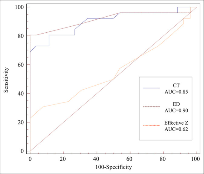
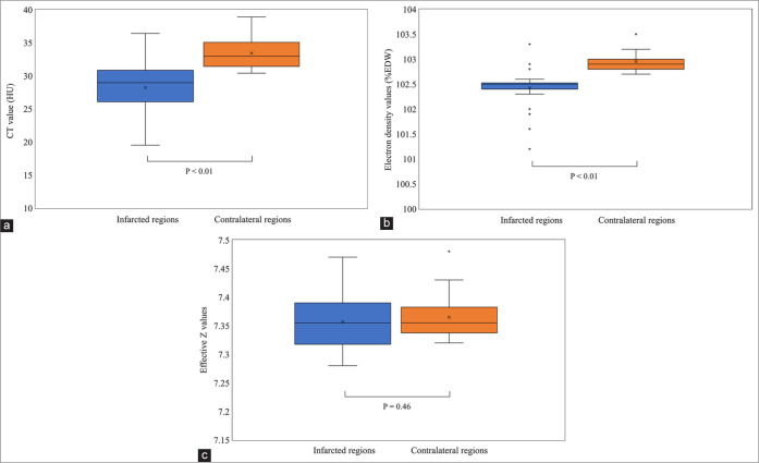
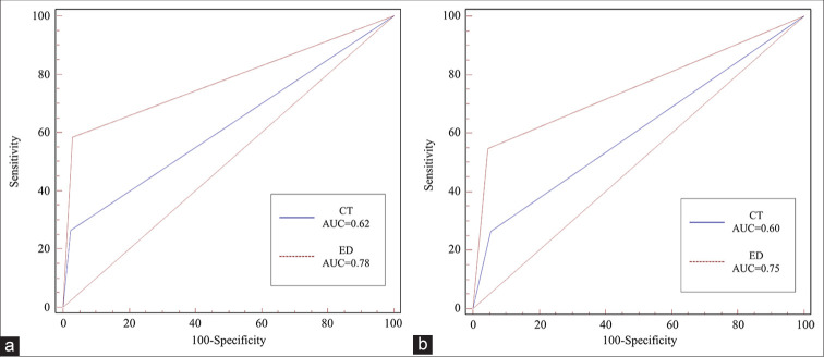
Results: Significant differences were observed in the conventional CT and ED values (P < 0.01) but not in effective Z values (P = 0.46) between the infarcted and contralateral regions. ED imaging demonstrated superior diagnostic accuracy (area under curve [AUC] = 0.90) compared with conventional 120-kVp CT (AUC = 0.85) and effective Z imaging (AUC = 0.62). Furthermore, interobserver agreement (kappa = 0.71) was better for ED imaging than for conventional 120-kVp CT (kappa = 0.65). Qualitative analysis revealed that ED images showed better agreement with MRI findings and higher interobserver consistency than conventional 120-kVp images.
Conclusion: Compared with conventional CT, DLCT with ED imaging significantly enhanced detection of EICs in AIS.
期刊介绍:
The Journal of Clinical Imaging Science (JCIS) is an open access peer-reviewed journal committed to publishing high-quality articles in the field of Imaging Science. The journal aims to present Imaging Science and relevant clinical information in an understandable and useful format. The journal is owned and published by the Scientific Scholar. Audience Our audience includes Radiologists, Researchers, Clinicians, medical professionals and students. Review process JCIS has a highly rigorous peer-review process that makes sure that manuscripts are scientifically accurate, relevant, novel and important. Authors disclose all conflicts, affiliations and financial associations such that the published content is not biased.

 求助内容:
求助内容: 应助结果提醒方式:


