Junhan Wei, Lei Zhang, Haiyan Wang, Qianfeng Wang, Wei Jia, Ru Wang, Runsheng Wang, Zhili Cui
{"title":"非动脉性急性缺血性视神经病变的结构-功能相关性。","authors":"Junhan Wei, Lei Zhang, Haiyan Wang, Qianfeng Wang, Wei Jia, Ru Wang, Runsheng Wang, Zhili Cui","doi":"10.2147/EB.S512882","DOIUrl":null,"url":null,"abstract":"<p><strong>Purpose: </strong>This study investigated the relationships between structural and functional parameters in non-arteritic ischemic optic neuropathy (NAION).</p><p><strong>Methods: </strong>This retrospective study enrolled 29 patients (58.2 ± 10.4 years old) with unilateral NAION. During the acute phase, we performed comprehensive evaluations including best-corrected visual acuity (BCVA), optical coherence tomography (OCT), optical coherence tomography angiography (OCTA), visual fields (VF), visual evoked potentials (VEP), electroretinography (ERG), and multifocal ERG (mf-ERG). At three months post-presentation, patients underwent follow-up assessments comprising visual acuity testing, perimetry, and advanced retinal imaging.</p><p><strong>Results: </strong>During the acute phase, affected eyes demonstrated increased mean retinal nerve fiber layer (RNFL) thickness, while ganglion cell-inner plexiform layer (GCIPL) thickness decreased. Both visual fields mean deviation (MD) and VEP P100 amplitude were reduced, accompanied by prolonged peak latency. We also observed decreased P1 response density in mf-ERG. Analysis revealed significant direct correlations between GCIPL parameters and electrophysiological measurements, particularly VEP P100 amplitude and mf-ERG P1 response density. Mean GCIPL thickness, VF MD, and VEP P100 amplitude showed negative correlations with baseline logMAR VA. Baseline VF MD, VEP P100 amplitude, and minimum GCIPL thickness showed negative correlations with logMAR VA at 3-month follow-up.</p><p><strong>Conclusion: </strong>Retinal ganglion cell layer thickness serves as a valuable indicator to objective evaluate optic nerve function in acute NAION patients. Decreases in both VEP amplitude and mf-ERG response density showed significant correlations with retinal ganglion cell layer thickness. Baseline visual field performance, VEP measurements, and minimum GCIPL thickness exhibited negative correlations visual acuity at 3-month follow-up.</p><p><strong>Trial registration: </strong>Clinical Research Ethics Committee of Xi'an People's Hospital (NO. 20220018). Registered 27 September 2022-Retrospectively registered, https://www.medicalresearch.org.cn/. Informed consent was obtained from each participant.</p>","PeriodicalId":51844,"journal":{"name":"Eye and Brain","volume":"17 ","pages":"13-25"},"PeriodicalIF":2.4000,"publicationDate":"2025-05-03","publicationTypes":"Journal Article","fieldsOfStudy":null,"isOpenAccess":false,"openAccessPdf":"https://www.ncbi.nlm.nih.gov/pmc/articles/PMC12057633/pdf/","citationCount":"0","resultStr":"{\"title\":\"Structural-Functional Correlation in Non-Arteritic Acute Ischemic Optic Neuropathy.\",\"authors\":\"Junhan Wei, Lei Zhang, Haiyan Wang, Qianfeng Wang, Wei Jia, Ru Wang, Runsheng Wang, Zhili Cui\",\"doi\":\"10.2147/EB.S512882\",\"DOIUrl\":null,\"url\":null,\"abstract\":\"<p><strong>Purpose: </strong>This study investigated the relationships between structural and functional parameters in non-arteritic ischemic optic neuropathy (NAION).</p><p><strong>Methods: </strong>This retrospective study enrolled 29 patients (58.2 ± 10.4 years old) with unilateral NAION. During the acute phase, we performed comprehensive evaluations including best-corrected visual acuity (BCVA), optical coherence tomography (OCT), optical coherence tomography angiography (OCTA), visual fields (VF), visual evoked potentials (VEP), electroretinography (ERG), and multifocal ERG (mf-ERG). At three months post-presentation, patients underwent follow-up assessments comprising visual acuity testing, perimetry, and advanced retinal imaging.</p><p><strong>Results: </strong>During the acute phase, affected eyes demonstrated increased mean retinal nerve fiber layer (RNFL) thickness, while ganglion cell-inner plexiform layer (GCIPL) thickness decreased. Both visual fields mean deviation (MD) and VEP P100 amplitude were reduced, accompanied by prolonged peak latency. We also observed decreased P1 response density in mf-ERG. Analysis revealed significant direct correlations between GCIPL parameters and electrophysiological measurements, particularly VEP P100 amplitude and mf-ERG P1 response density. Mean GCIPL thickness, VF MD, and VEP P100 amplitude showed negative correlations with baseline logMAR VA. Baseline VF MD, VEP P100 amplitude, and minimum GCIPL thickness showed negative correlations with logMAR VA at 3-month follow-up.</p><p><strong>Conclusion: </strong>Retinal ganglion cell layer thickness serves as a valuable indicator to objective evaluate optic nerve function in acute NAION patients. Decreases in both VEP amplitude and mf-ERG response density showed significant correlations with retinal ganglion cell layer thickness. Baseline visual field performance, VEP measurements, and minimum GCIPL thickness exhibited negative correlations visual acuity at 3-month follow-up.</p><p><strong>Trial registration: </strong>Clinical Research Ethics Committee of Xi'an People's Hospital (NO. 20220018). Registered 27 September 2022-Retrospectively registered, https://www.medicalresearch.org.cn/. Informed consent was obtained from each participant.</p>\",\"PeriodicalId\":51844,\"journal\":{\"name\":\"Eye and Brain\",\"volume\":\"17 \",\"pages\":\"13-25\"},\"PeriodicalIF\":2.4000,\"publicationDate\":\"2025-05-03\",\"publicationTypes\":\"Journal Article\",\"fieldsOfStudy\":null,\"isOpenAccess\":false,\"openAccessPdf\":\"https://www.ncbi.nlm.nih.gov/pmc/articles/PMC12057633/pdf/\",\"citationCount\":\"0\",\"resultStr\":null,\"platform\":\"Semanticscholar\",\"paperid\":null,\"PeriodicalName\":\"Eye and Brain\",\"FirstCategoryId\":\"1085\",\"ListUrlMain\":\"https://doi.org/10.2147/EB.S512882\",\"RegionNum\":0,\"RegionCategory\":null,\"ArticlePicture\":[],\"TitleCN\":null,\"AbstractTextCN\":null,\"PMCID\":null,\"EPubDate\":\"2025/1/1 0:00:00\",\"PubModel\":\"eCollection\",\"JCR\":\"Q1\",\"JCRName\":\"OPHTHALMOLOGY\",\"Score\":null,\"Total\":0}","platform":"Semanticscholar","paperid":null,"PeriodicalName":"Eye and Brain","FirstCategoryId":"1085","ListUrlMain":"https://doi.org/10.2147/EB.S512882","RegionNum":0,"RegionCategory":null,"ArticlePicture":[],"TitleCN":null,"AbstractTextCN":null,"PMCID":null,"EPubDate":"2025/1/1 0:00:00","PubModel":"eCollection","JCR":"Q1","JCRName":"OPHTHALMOLOGY","Score":null,"Total":0}
Structural-Functional Correlation in Non-Arteritic Acute Ischemic Optic Neuropathy.
Purpose: This study investigated the relationships between structural and functional parameters in non-arteritic ischemic optic neuropathy (NAION).
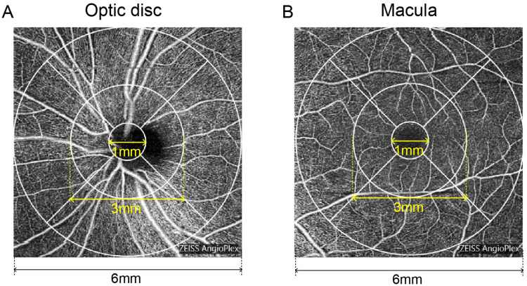
Methods: This retrospective study enrolled 29 patients (58.2 ± 10.4 years old) with unilateral NAION. During the acute phase, we performed comprehensive evaluations including best-corrected visual acuity (BCVA), optical coherence tomography (OCT), optical coherence tomography angiography (OCTA), visual fields (VF), visual evoked potentials (VEP), electroretinography (ERG), and multifocal ERG (mf-ERG). At three months post-presentation, patients underwent follow-up assessments comprising visual acuity testing, perimetry, and advanced retinal imaging.
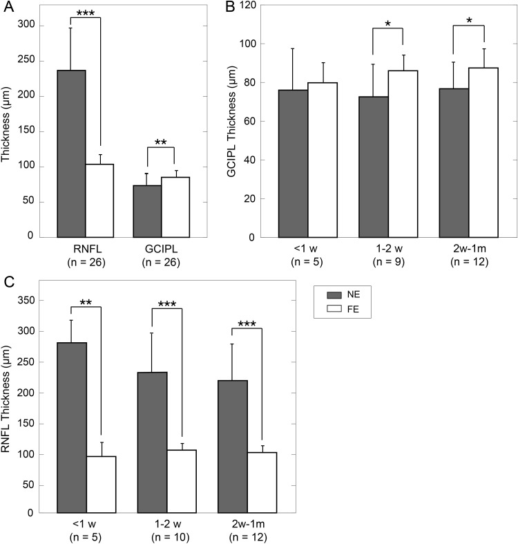
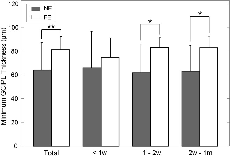
Results: During the acute phase, affected eyes demonstrated increased mean retinal nerve fiber layer (RNFL) thickness, while ganglion cell-inner plexiform layer (GCIPL) thickness decreased. Both visual fields mean deviation (MD) and VEP P100 amplitude were reduced, accompanied by prolonged peak latency. We also observed decreased P1 response density in mf-ERG. Analysis revealed significant direct correlations between GCIPL parameters and electrophysiological measurements, particularly VEP P100 amplitude and mf-ERG P1 response density. Mean GCIPL thickness, VF MD, and VEP P100 amplitude showed negative correlations with baseline logMAR VA. Baseline VF MD, VEP P100 amplitude, and minimum GCIPL thickness showed negative correlations with logMAR VA at 3-month follow-up.
Conclusion: Retinal ganglion cell layer thickness serves as a valuable indicator to objective evaluate optic nerve function in acute NAION patients. Decreases in both VEP amplitude and mf-ERG response density showed significant correlations with retinal ganglion cell layer thickness. Baseline visual field performance, VEP measurements, and minimum GCIPL thickness exhibited negative correlations visual acuity at 3-month follow-up.
Trial registration: Clinical Research Ethics Committee of Xi'an People's Hospital (NO. 20220018). Registered 27 September 2022-Retrospectively registered, https://www.medicalresearch.org.cn/. Informed consent was obtained from each participant.
期刊介绍:
Eye and Brain is an international, peer-reviewed, open access journal focusing on basic research, clinical findings, and expert reviews in the field of visual science and neuro-ophthalmology. The journal’s unique focus is the link between two well-known visual centres, the eye and the brain, with an emphasis on the importance of such connections. All aspects of clinical and especially basic research on the visual system are addressed within the journal as well as significant future directions in vision research and therapeutic measures. This unique journal focuses on neurological aspects of vision – both physiological and pathological. The scope of the journal spans from the cornea to the associational visual cortex and all the visual centers in between. Topics range from basic biological mechanisms to therapeutic treatment, from simple organisms to humans, and utilizing techniques from molecular biology to behavior. The journal especially welcomes primary research articles or review papers that make the connection between the eye and the brain. Specific areas covered in the journal include: Physiology and pathophysiology of visual centers, Eye movement disorders and strabismus, Cellular, biochemical, and molecular features of the visual system, Structural and functional organization of the eye and of the visual cortex, Metabolic demands of the visual system, Diseases and disorders with neuro-ophthalmic manifestations, Clinical and experimental neuro-ophthalmology and visual system pathologies, Epidemiological studies.

 求助内容:
求助内容: 应助结果提醒方式:


