{"title":"当前和未来的作用和先进的CT在炎性关节炎的临床和试验中的应用。","authors":"Torsten Diekhoff, Sevtap Tugce Ulas","doi":"10.1007/s00256-025-04931-4","DOIUrl":null,"url":null,"abstract":"<p><p>Computed tomography (CT) has traditionally been underutilized in the imaging of inflammatory arthritis due to its limitations in assessing soft tissue inflammation and concerns over radiation exposure. However, recent technological advancements have positioned CT as a more viable imaging modality for arthritis, offering high specificity and sensitivity in detecting structural bone changes. However, advances in ultra-low-dose CT protocols and AI-driven image reconstruction have significantly reduced radiation exposure while maintaining diagnostic quality. Dynamic CT and spectral CT techniques, including dual-energy CT (DECT), have broadened CT's application in assessing dynamic joint instabilities and visualizing inflammatory changes through material-specific imaging. Techniques such as CT subtraction imaging and iodine mapping have enhanced the detection of active soft-tissue inflammation, virtual non-calcium reconstructions, and the detection of bone marrow edema. Possible CT applications span various forms of arthritis, including gout, calcium pyrophosphate deposition disease (CPPD), psoriatic arthritis, and axial spondyloarthritis. Beyond its diagnostic capabilities, CT's ability to provide detailed structural assessment positions is a valuable tool for monitoring disease progression and therapeutic response, particularly in clinical trials. While MRI remains superior for soft tissue evaluation, CT's specificity for bone-related changes and its potential for integration into routine arthritis management warrant further exploration and research. This review explores the current and emerging roles of CT in arthritis diagnostics, with a focus on novel applications and future potential.</p>","PeriodicalId":21783,"journal":{"name":"Skeletal Radiology","volume":" ","pages":"2385-2397"},"PeriodicalIF":2.2000,"publicationDate":"2025-11-01","publicationTypes":"Journal Article","fieldsOfStudy":null,"isOpenAccess":false,"openAccessPdf":"https://www.ncbi.nlm.nih.gov/pmc/articles/PMC12460486/pdf/","citationCount":"0","resultStr":"{\"title\":\"Current and future role of CT and advanced CT applications in inflammatory arthritis in the clinic and trials.\",\"authors\":\"Torsten Diekhoff, Sevtap Tugce Ulas\",\"doi\":\"10.1007/s00256-025-04931-4\",\"DOIUrl\":null,\"url\":null,\"abstract\":\"<p><p>Computed tomography (CT) has traditionally been underutilized in the imaging of inflammatory arthritis due to its limitations in assessing soft tissue inflammation and concerns over radiation exposure. However, recent technological advancements have positioned CT as a more viable imaging modality for arthritis, offering high specificity and sensitivity in detecting structural bone changes. However, advances in ultra-low-dose CT protocols and AI-driven image reconstruction have significantly reduced radiation exposure while maintaining diagnostic quality. Dynamic CT and spectral CT techniques, including dual-energy CT (DECT), have broadened CT's application in assessing dynamic joint instabilities and visualizing inflammatory changes through material-specific imaging. Techniques such as CT subtraction imaging and iodine mapping have enhanced the detection of active soft-tissue inflammation, virtual non-calcium reconstructions, and the detection of bone marrow edema. Possible CT applications span various forms of arthritis, including gout, calcium pyrophosphate deposition disease (CPPD), psoriatic arthritis, and axial spondyloarthritis. Beyond its diagnostic capabilities, CT's ability to provide detailed structural assessment positions is a valuable tool for monitoring disease progression and therapeutic response, particularly in clinical trials. While MRI remains superior for soft tissue evaluation, CT's specificity for bone-related changes and its potential for integration into routine arthritis management warrant further exploration and research. This review explores the current and emerging roles of CT in arthritis diagnostics, with a focus on novel applications and future potential.</p>\",\"PeriodicalId\":21783,\"journal\":{\"name\":\"Skeletal Radiology\",\"volume\":\" \",\"pages\":\"2385-2397\"},\"PeriodicalIF\":2.2000,\"publicationDate\":\"2025-11-01\",\"publicationTypes\":\"Journal Article\",\"fieldsOfStudy\":null,\"isOpenAccess\":false,\"openAccessPdf\":\"https://www.ncbi.nlm.nih.gov/pmc/articles/PMC12460486/pdf/\",\"citationCount\":\"0\",\"resultStr\":null,\"platform\":\"Semanticscholar\",\"paperid\":null,\"PeriodicalName\":\"Skeletal Radiology\",\"FirstCategoryId\":\"3\",\"ListUrlMain\":\"https://doi.org/10.1007/s00256-025-04931-4\",\"RegionNum\":3,\"RegionCategory\":\"医学\",\"ArticlePicture\":[],\"TitleCN\":null,\"AbstractTextCN\":null,\"PMCID\":null,\"EPubDate\":\"2025/4/16 0:00:00\",\"PubModel\":\"Epub\",\"JCR\":\"Q2\",\"JCRName\":\"ORTHOPEDICS\",\"Score\":null,\"Total\":0}","platform":"Semanticscholar","paperid":null,"PeriodicalName":"Skeletal Radiology","FirstCategoryId":"3","ListUrlMain":"https://doi.org/10.1007/s00256-025-04931-4","RegionNum":3,"RegionCategory":"医学","ArticlePicture":[],"TitleCN":null,"AbstractTextCN":null,"PMCID":null,"EPubDate":"2025/4/16 0:00:00","PubModel":"Epub","JCR":"Q2","JCRName":"ORTHOPEDICS","Score":null,"Total":0}
Current and future role of CT and advanced CT applications in inflammatory arthritis in the clinic and trials.
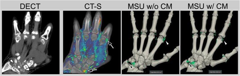
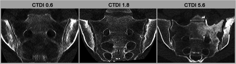
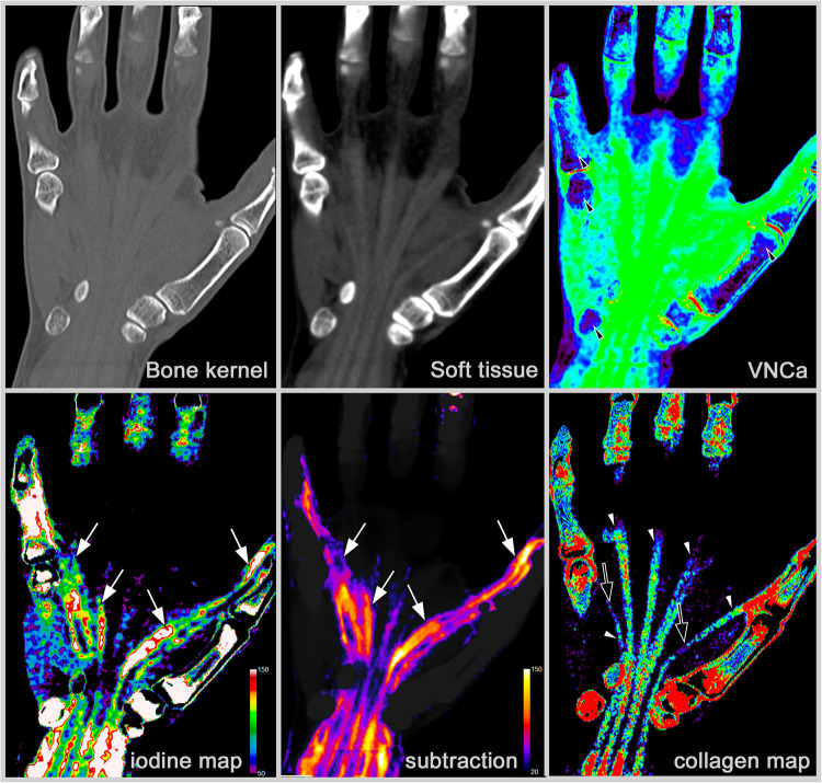
Computed tomography (CT) has traditionally been underutilized in the imaging of inflammatory arthritis due to its limitations in assessing soft tissue inflammation and concerns over radiation exposure. However, recent technological advancements have positioned CT as a more viable imaging modality for arthritis, offering high specificity and sensitivity in detecting structural bone changes. However, advances in ultra-low-dose CT protocols and AI-driven image reconstruction have significantly reduced radiation exposure while maintaining diagnostic quality. Dynamic CT and spectral CT techniques, including dual-energy CT (DECT), have broadened CT's application in assessing dynamic joint instabilities and visualizing inflammatory changes through material-specific imaging. Techniques such as CT subtraction imaging and iodine mapping have enhanced the detection of active soft-tissue inflammation, virtual non-calcium reconstructions, and the detection of bone marrow edema. Possible CT applications span various forms of arthritis, including gout, calcium pyrophosphate deposition disease (CPPD), psoriatic arthritis, and axial spondyloarthritis. Beyond its diagnostic capabilities, CT's ability to provide detailed structural assessment positions is a valuable tool for monitoring disease progression and therapeutic response, particularly in clinical trials. While MRI remains superior for soft tissue evaluation, CT's specificity for bone-related changes and its potential for integration into routine arthritis management warrant further exploration and research. This review explores the current and emerging roles of CT in arthritis diagnostics, with a focus on novel applications and future potential.
期刊介绍:
Skeletal Radiology provides a forum for the dissemination of current knowledge and information dealing with disorders of the musculoskeletal system including the spine. While emphasizing the radiological aspects of the many varied skeletal abnormalities, the journal also adopts an interdisciplinary approach, reflecting the membership of the International Skeletal Society. Thus, the anatomical, pathological, physiological, clinical, metabolic and epidemiological aspects of the many entities affecting the skeleton receive appropriate consideration.
This is the Journal of the International Skeletal Society and the Official Journal of the Society of Skeletal Radiology and the Australasian Musculoskelelal Imaging Group.

 求助内容:
求助内容: 应助结果提醒方式:


