{"title":"肥胖对肥厚性心肌病患者心肌组织特征的影响:一项基于心血管磁共振的研究","authors":"Jie Wang, Lutong Pu, Jinquan Zhang, Ruihao Xu, Yang Li, Mengdi Yu, Yangjie Li, Jiajun Guo, Yuanwei Xu, Yu Kang, Yuchi Han, Yucheng Chen","doi":"10.1016/j.jocmr.2025.101903","DOIUrl":null,"url":null,"abstract":"<p><strong>Background: </strong>Obesity is associated with cardiac steatosis in healthy adults and is independently associated with increased left ventricular (LV) mass and could contribute to the progression of heart failure-related composite events in patients with hypertrophic cardiomyopathy (HCM). However, it is unclear whether the increased LV mass is accompanied by increased fibrosis. We aimed to assess the impact of increased body mass index (BMI) on myocardial tissue characteristics in patients with HCM.</p><p><strong>Methods: </strong>A total of 737 patients with HCM (99 obese, 298 overweight, and 340 normal-weight patients) who underwent cardiovascular magnetic resonance (CMR) imaging were prospectively included. We assessed the relationship between BMI and LV mass, global native T1, extracellular volume, and late gadolinium enhancement (LGE) using CMR. Myocardial tissues from one patient each with obstructive HCM who underwent septal myectomy of the obese, overweight, and normal-weight groups were obtained and stained with red oil O, hematoxylin, and Masson's trichrome.</p><p><strong>Results: </strong>LV mass index (87.2, interquartile range [IQR]: 71.3 to 113.8, 89.4, IQR:75.5 to 111.5, and 104.7, IQR: 86.4 to 123.4 g/m<sup>2</sup>, P < 0.001) was higher in obese and overweight patients with HCM than those with normal weight, but the native T1 was decreased in obese patients with HCM (1324±67 ms, 1308±63 ms, and 1298±67 ms, P < 0.001). In addition, there was no significant difference in LGE extent among the three subgroups (normal weight: 3.7%, IQR: 0 to 9.5%, overweight: 2.7%, IQR: 0 to 7.7%, obese: 3.8%, IQR: 0 to 7.2%, P = 0.194). Multivariable linear regression analyses found that BMI was independently associated with global native T1 (β = -1.918, P = 0.005). Furthermore, myocardial tissues stained with oil red O from three patients showed an increasing extent of fat deposits with BMI, whereas collagen volume fractions were similar.</p><p><strong>Conclusion: </strong>In HCM patients, obesity is associated with increased myocardial mass and decreased native T1, likely reflecting cardiac steatosis in addition to fibrosis. This distinction underscores the potential reversibility of obesity-related myocardial changes through targeted weight management.</p><p><strong>Trial registration: </strong>This prospective cohort study was registered in the Chinese Clinical Trial Registry (URL: http://www.chictr.org.cn; Registry number: ChiCTR1900024094).</p>","PeriodicalId":15221,"journal":{"name":"Journal of Cardiovascular Magnetic Resonance","volume":" ","pages":"101903"},"PeriodicalIF":6.1000,"publicationDate":"2025-01-01","publicationTypes":"Journal Article","fieldsOfStudy":null,"isOpenAccess":false,"openAccessPdf":"https://www.ncbi.nlm.nih.gov/pmc/articles/PMC12141553/pdf/","citationCount":"0","resultStr":"{\"title\":\"Effect of obesity on myocardial tissue characteristics in patients with hypertrophic cardiomyopathy: a cardiovascular magnetic resonance-based study.\",\"authors\":\"Jie Wang, Lutong Pu, Jinquan Zhang, Ruihao Xu, Yang Li, Mengdi Yu, Yangjie Li, Jiajun Guo, Yuanwei Xu, Yu Kang, Yuchi Han, Yucheng Chen\",\"doi\":\"10.1016/j.jocmr.2025.101903\",\"DOIUrl\":null,\"url\":null,\"abstract\":\"<p><strong>Background: </strong>Obesity is associated with cardiac steatosis in healthy adults and is independently associated with increased left ventricular (LV) mass and could contribute to the progression of heart failure-related composite events in patients with hypertrophic cardiomyopathy (HCM). However, it is unclear whether the increased LV mass is accompanied by increased fibrosis. We aimed to assess the impact of increased body mass index (BMI) on myocardial tissue characteristics in patients with HCM.</p><p><strong>Methods: </strong>A total of 737 patients with HCM (99 obese, 298 overweight, and 340 normal-weight patients) who underwent cardiovascular magnetic resonance (CMR) imaging were prospectively included. We assessed the relationship between BMI and LV mass, global native T1, extracellular volume, and late gadolinium enhancement (LGE) using CMR. Myocardial tissues from one patient each with obstructive HCM who underwent septal myectomy of the obese, overweight, and normal-weight groups were obtained and stained with red oil O, hematoxylin, and Masson's trichrome.</p><p><strong>Results: </strong>LV mass index (87.2, interquartile range [IQR]: 71.3 to 113.8, 89.4, IQR:75.5 to 111.5, and 104.7, IQR: 86.4 to 123.4 g/m<sup>2</sup>, P < 0.001) was higher in obese and overweight patients with HCM than those with normal weight, but the native T1 was decreased in obese patients with HCM (1324±67 ms, 1308±63 ms, and 1298±67 ms, P < 0.001). In addition, there was no significant difference in LGE extent among the three subgroups (normal weight: 3.7%, IQR: 0 to 9.5%, overweight: 2.7%, IQR: 0 to 7.7%, obese: 3.8%, IQR: 0 to 7.2%, P = 0.194). Multivariable linear regression analyses found that BMI was independently associated with global native T1 (β = -1.918, P = 0.005). Furthermore, myocardial tissues stained with oil red O from three patients showed an increasing extent of fat deposits with BMI, whereas collagen volume fractions were similar.</p><p><strong>Conclusion: </strong>In HCM patients, obesity is associated with increased myocardial mass and decreased native T1, likely reflecting cardiac steatosis in addition to fibrosis. This distinction underscores the potential reversibility of obesity-related myocardial changes through targeted weight management.</p><p><strong>Trial registration: </strong>This prospective cohort study was registered in the Chinese Clinical Trial Registry (URL: http://www.chictr.org.cn; Registry number: ChiCTR1900024094).</p>\",\"PeriodicalId\":15221,\"journal\":{\"name\":\"Journal of Cardiovascular Magnetic Resonance\",\"volume\":\" \",\"pages\":\"101903\"},\"PeriodicalIF\":6.1000,\"publicationDate\":\"2025-01-01\",\"publicationTypes\":\"Journal Article\",\"fieldsOfStudy\":null,\"isOpenAccess\":false,\"openAccessPdf\":\"https://www.ncbi.nlm.nih.gov/pmc/articles/PMC12141553/pdf/\",\"citationCount\":\"0\",\"resultStr\":null,\"platform\":\"Semanticscholar\",\"paperid\":null,\"PeriodicalName\":\"Journal of Cardiovascular Magnetic Resonance\",\"FirstCategoryId\":\"3\",\"ListUrlMain\":\"https://doi.org/10.1016/j.jocmr.2025.101903\",\"RegionNum\":1,\"RegionCategory\":\"医学\",\"ArticlePicture\":[],\"TitleCN\":null,\"AbstractTextCN\":null,\"PMCID\":null,\"EPubDate\":\"2025/5/2 0:00:00\",\"PubModel\":\"Epub\",\"JCR\":\"Q1\",\"JCRName\":\"CARDIAC & CARDIOVASCULAR SYSTEMS\",\"Score\":null,\"Total\":0}","platform":"Semanticscholar","paperid":null,"PeriodicalName":"Journal of Cardiovascular Magnetic Resonance","FirstCategoryId":"3","ListUrlMain":"https://doi.org/10.1016/j.jocmr.2025.101903","RegionNum":1,"RegionCategory":"医学","ArticlePicture":[],"TitleCN":null,"AbstractTextCN":null,"PMCID":null,"EPubDate":"2025/5/2 0:00:00","PubModel":"Epub","JCR":"Q1","JCRName":"CARDIAC & CARDIOVASCULAR SYSTEMS","Score":null,"Total":0}
Effect of obesity on myocardial tissue characteristics in patients with hypertrophic cardiomyopathy: a cardiovascular magnetic resonance-based study.
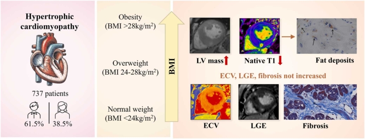
Background: Obesity is associated with cardiac steatosis in healthy adults and is independently associated with increased left ventricular (LV) mass and could contribute to the progression of heart failure-related composite events in patients with hypertrophic cardiomyopathy (HCM). However, it is unclear whether the increased LV mass is accompanied by increased fibrosis. We aimed to assess the impact of increased body mass index (BMI) on myocardial tissue characteristics in patients with HCM.
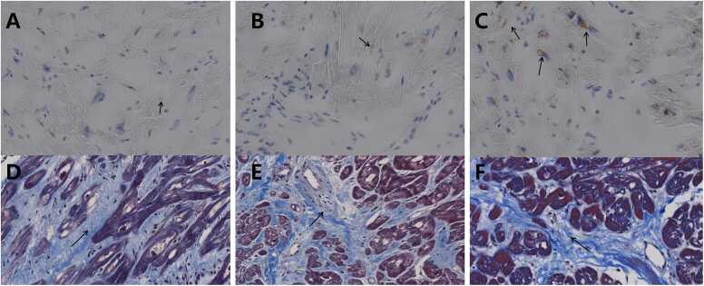
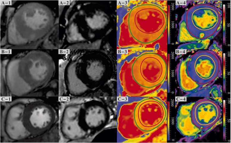
Methods: A total of 737 patients with HCM (99 obese, 298 overweight, and 340 normal-weight patients) who underwent cardiovascular magnetic resonance (CMR) imaging were prospectively included. We assessed the relationship between BMI and LV mass, global native T1, extracellular volume, and late gadolinium enhancement (LGE) using CMR. Myocardial tissues from one patient each with obstructive HCM who underwent septal myectomy of the obese, overweight, and normal-weight groups were obtained and stained with red oil O, hematoxylin, and Masson's trichrome.
Results: LV mass index (87.2, interquartile range [IQR]: 71.3 to 113.8, 89.4, IQR:75.5 to 111.5, and 104.7, IQR: 86.4 to 123.4 g/m2, P < 0.001) was higher in obese and overweight patients with HCM than those with normal weight, but the native T1 was decreased in obese patients with HCM (1324±67 ms, 1308±63 ms, and 1298±67 ms, P < 0.001). In addition, there was no significant difference in LGE extent among the three subgroups (normal weight: 3.7%, IQR: 0 to 9.5%, overweight: 2.7%, IQR: 0 to 7.7%, obese: 3.8%, IQR: 0 to 7.2%, P = 0.194). Multivariable linear regression analyses found that BMI was independently associated with global native T1 (β = -1.918, P = 0.005). Furthermore, myocardial tissues stained with oil red O from three patients showed an increasing extent of fat deposits with BMI, whereas collagen volume fractions were similar.
Conclusion: In HCM patients, obesity is associated with increased myocardial mass and decreased native T1, likely reflecting cardiac steatosis in addition to fibrosis. This distinction underscores the potential reversibility of obesity-related myocardial changes through targeted weight management.
Trial registration: This prospective cohort study was registered in the Chinese Clinical Trial Registry (URL: http://www.chictr.org.cn; Registry number: ChiCTR1900024094).
期刊介绍:
Journal of Cardiovascular Magnetic Resonance (JCMR) publishes high-quality articles on all aspects of basic, translational and clinical research on the design, development, manufacture, and evaluation of cardiovascular magnetic resonance (CMR) methods applied to the cardiovascular system. Topical areas include, but are not limited to:
New applications of magnetic resonance to improve the diagnostic strategies, risk stratification, characterization and management of diseases affecting the cardiovascular system.
New methods to enhance or accelerate image acquisition and data analysis.
Results of multicenter, or larger single-center studies that provide insight into the utility of CMR.
Basic biological perceptions derived by CMR methods.

 求助内容:
求助内容: 应助结果提醒方式:


