{"title":"猫腹壁球囊瘤。","authors":"Lisa Castellano, Debra Fews","doi":"10.1177/20551169251332395","DOIUrl":null,"url":null,"abstract":"<p><p>Case summaryA 4-year-old male castrated domestic shorthair cat presented with a 2 cm painful cutaneous mass on its right lateral abdominal wall. The cat inflicted self-trauma to the lesion site causing secondary ulceration and mild haemorrhage. Fine-needle aspiration or incisional biopsy was advised, along with diagnostic imaging; however, excisional biopsy was preferred by the owners. The mass was surgically removed with 1 cm lateral margins and a deep fascial plane. The histopathological features were most consistent with a diagnosis of a glomus tumour; the diagnosis was supported by subsequent immunohistochemistry. The tumour was completely removed and there were no signs of recurrence at the 6-month follow-up. In this case, surgery is expected to be curative. <i>Relevance and novel information</i> To the authors' knowledge, this is the first report of a glomus tumour of the body wall in a cat and its association with pain and self-trauma. This report aims to add more data to the diagnosis and presentation of glomus tumours in animals.</p>","PeriodicalId":36588,"journal":{"name":"Journal of Feline Medicine and Surgery Open Reports","volume":"11 1","pages":"20551169251332395"},"PeriodicalIF":0.7000,"publicationDate":"2025-05-07","publicationTypes":"Journal Article","fieldsOfStudy":null,"isOpenAccess":false,"openAccessPdf":"https://www.ncbi.nlm.nih.gov/pmc/articles/PMC12062611/pdf/","citationCount":"0","resultStr":"{\"title\":\"Glomus tumour of the abdominal wall in a cat.\",\"authors\":\"Lisa Castellano, Debra Fews\",\"doi\":\"10.1177/20551169251332395\",\"DOIUrl\":null,\"url\":null,\"abstract\":\"<p><p>Case summaryA 4-year-old male castrated domestic shorthair cat presented with a 2 cm painful cutaneous mass on its right lateral abdominal wall. The cat inflicted self-trauma to the lesion site causing secondary ulceration and mild haemorrhage. Fine-needle aspiration or incisional biopsy was advised, along with diagnostic imaging; however, excisional biopsy was preferred by the owners. The mass was surgically removed with 1 cm lateral margins and a deep fascial plane. The histopathological features were most consistent with a diagnosis of a glomus tumour; the diagnosis was supported by subsequent immunohistochemistry. The tumour was completely removed and there were no signs of recurrence at the 6-month follow-up. In this case, surgery is expected to be curative. <i>Relevance and novel information</i> To the authors' knowledge, this is the first report of a glomus tumour of the body wall in a cat and its association with pain and self-trauma. This report aims to add more data to the diagnosis and presentation of glomus tumours in animals.</p>\",\"PeriodicalId\":36588,\"journal\":{\"name\":\"Journal of Feline Medicine and Surgery Open Reports\",\"volume\":\"11 1\",\"pages\":\"20551169251332395\"},\"PeriodicalIF\":0.7000,\"publicationDate\":\"2025-05-07\",\"publicationTypes\":\"Journal Article\",\"fieldsOfStudy\":null,\"isOpenAccess\":false,\"openAccessPdf\":\"https://www.ncbi.nlm.nih.gov/pmc/articles/PMC12062611/pdf/\",\"citationCount\":\"0\",\"resultStr\":null,\"platform\":\"Semanticscholar\",\"paperid\":null,\"PeriodicalName\":\"Journal of Feline Medicine and Surgery Open Reports\",\"FirstCategoryId\":\"1085\",\"ListUrlMain\":\"https://doi.org/10.1177/20551169251332395\",\"RegionNum\":0,\"RegionCategory\":null,\"ArticlePicture\":[],\"TitleCN\":null,\"AbstractTextCN\":null,\"PMCID\":null,\"EPubDate\":\"2025/1/1 0:00:00\",\"PubModel\":\"eCollection\",\"JCR\":\"Q3\",\"JCRName\":\"VETERINARY SCIENCES\",\"Score\":null,\"Total\":0}","platform":"Semanticscholar","paperid":null,"PeriodicalName":"Journal of Feline Medicine and Surgery Open Reports","FirstCategoryId":"1085","ListUrlMain":"https://doi.org/10.1177/20551169251332395","RegionNum":0,"RegionCategory":null,"ArticlePicture":[],"TitleCN":null,"AbstractTextCN":null,"PMCID":null,"EPubDate":"2025/1/1 0:00:00","PubModel":"eCollection","JCR":"Q3","JCRName":"VETERINARY SCIENCES","Score":null,"Total":0}
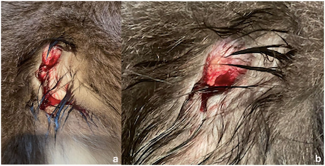
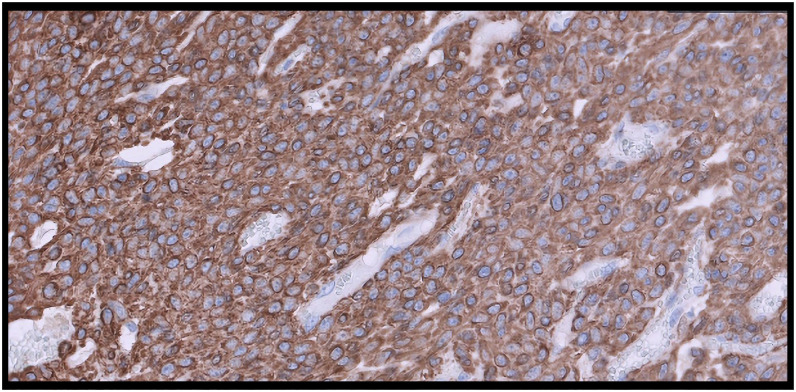
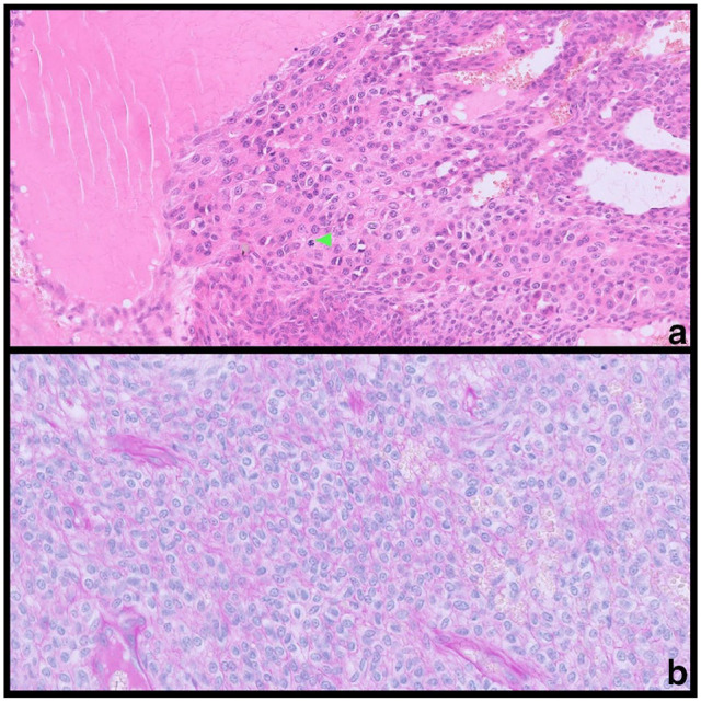
Case summaryA 4-year-old male castrated domestic shorthair cat presented with a 2 cm painful cutaneous mass on its right lateral abdominal wall. The cat inflicted self-trauma to the lesion site causing secondary ulceration and mild haemorrhage. Fine-needle aspiration or incisional biopsy was advised, along with diagnostic imaging; however, excisional biopsy was preferred by the owners. The mass was surgically removed with 1 cm lateral margins and a deep fascial plane. The histopathological features were most consistent with a diagnosis of a glomus tumour; the diagnosis was supported by subsequent immunohistochemistry. The tumour was completely removed and there were no signs of recurrence at the 6-month follow-up. In this case, surgery is expected to be curative. Relevance and novel information To the authors' knowledge, this is the first report of a glomus tumour of the body wall in a cat and its association with pain and self-trauma. This report aims to add more data to the diagnosis and presentation of glomus tumours in animals.

 求助内容:
求助内容: 应助结果提醒方式:


