{"title":"乳腺细针抽吸器的横滨分类及其与组织病理学的相关性。","authors":"Vishal Rohilla, Parveen Kundu, Monika Gathwal, Pushpendra Malik, Chiranjeev Gathwal, Sunaina Hooda","doi":"10.4274/ejbh.galenos.2025.2024-11-4","DOIUrl":null,"url":null,"abstract":"<p><strong>Objective: </strong>Breast cancer is the most prevalent cancer among women worldwide. In developing countries, fine needle aspiration cytology (FNAC) is commonly used for screening to reduce mortality rates. The International Academy of Cytology has established the Yokohama system to enhance diagnostic clarity and communication between pathologists and clinicians. A triple test approach, incorporating clinical evaluation, imaging, and FNAC, can further improve patient care for breast lesions and may enhance the Yokohama System's effectiveness.</p><p><strong>Materials and methods: </strong>A prospective study about breast FNAC was done over a period of one year, from October 2022 to September 2023. The study involved patients with breast lesion referred for FNAC in the department of Pathology. The FNAC results were further classified using the Yokohama system for reporting breast cytopathology, 2016. The cytological findings were correlated with available histopathological results.</p><p><strong>Results: </strong>In the study of 104 cases, 60 (57.7%) of whom had available histopathology results, breast lesions were categorized using the Yokohama system as: 7.7% insufficient, 47.1% benign, 26.9% atypical, 2.9% suspicious of malignancy, and 15.4% malignant. The risk of malignancy varied by category: 0% for category 1, 3.2% for category 2, 47% for category 3, and 100% for categories 4 and 5. The maximum sensitivity was 94.7% when considering atypical, suspicious, and malignant cases as positive. The highest specificity was 97.56% for malignant cases alone, while the best diagnostic accuracy was 83.3% when both malignant and suspicious cases were counted as positive.</p><p><strong>Conclusion: </strong>The Yokohama system effectively classified borderline lesions, facilitating early detection and improved management options. By integrating FNAC with standardized reporting, healthcare providers can make informed decisions, enhancing the diagnosis and treatment of breast lesions.</p>","PeriodicalId":93996,"journal":{"name":"European journal of breast health","volume":" ","pages":"237-245"},"PeriodicalIF":1.7000,"publicationDate":"2025-06-20","publicationTypes":"Journal Article","fieldsOfStudy":null,"isOpenAccess":false,"openAccessPdf":"https://www.ncbi.nlm.nih.gov/pmc/articles/PMC12180108/pdf/","citationCount":"0","resultStr":"{\"title\":\"Categorization of Breast Fine Needle Aspirates Using Yokohama Classification and Its Correlation With Histopathological Findings.\",\"authors\":\"Vishal Rohilla, Parveen Kundu, Monika Gathwal, Pushpendra Malik, Chiranjeev Gathwal, Sunaina Hooda\",\"doi\":\"10.4274/ejbh.galenos.2025.2024-11-4\",\"DOIUrl\":null,\"url\":null,\"abstract\":\"<p><strong>Objective: </strong>Breast cancer is the most prevalent cancer among women worldwide. In developing countries, fine needle aspiration cytology (FNAC) is commonly used for screening to reduce mortality rates. The International Academy of Cytology has established the Yokohama system to enhance diagnostic clarity and communication between pathologists and clinicians. A triple test approach, incorporating clinical evaluation, imaging, and FNAC, can further improve patient care for breast lesions and may enhance the Yokohama System's effectiveness.</p><p><strong>Materials and methods: </strong>A prospective study about breast FNAC was done over a period of one year, from October 2022 to September 2023. The study involved patients with breast lesion referred for FNAC in the department of Pathology. The FNAC results were further classified using the Yokohama system for reporting breast cytopathology, 2016. The cytological findings were correlated with available histopathological results.</p><p><strong>Results: </strong>In the study of 104 cases, 60 (57.7%) of whom had available histopathology results, breast lesions were categorized using the Yokohama system as: 7.7% insufficient, 47.1% benign, 26.9% atypical, 2.9% suspicious of malignancy, and 15.4% malignant. The risk of malignancy varied by category: 0% for category 1, 3.2% for category 2, 47% for category 3, and 100% for categories 4 and 5. The maximum sensitivity was 94.7% when considering atypical, suspicious, and malignant cases as positive. The highest specificity was 97.56% for malignant cases alone, while the best diagnostic accuracy was 83.3% when both malignant and suspicious cases were counted as positive.</p><p><strong>Conclusion: </strong>The Yokohama system effectively classified borderline lesions, facilitating early detection and improved management options. By integrating FNAC with standardized reporting, healthcare providers can make informed decisions, enhancing the diagnosis and treatment of breast lesions.</p>\",\"PeriodicalId\":93996,\"journal\":{\"name\":\"European journal of breast health\",\"volume\":\" \",\"pages\":\"237-245\"},\"PeriodicalIF\":1.7000,\"publicationDate\":\"2025-06-20\",\"publicationTypes\":\"Journal Article\",\"fieldsOfStudy\":null,\"isOpenAccess\":false,\"openAccessPdf\":\"https://www.ncbi.nlm.nih.gov/pmc/articles/PMC12180108/pdf/\",\"citationCount\":\"0\",\"resultStr\":null,\"platform\":\"Semanticscholar\",\"paperid\":null,\"PeriodicalName\":\"European journal of breast health\",\"FirstCategoryId\":\"1085\",\"ListUrlMain\":\"https://doi.org/10.4274/ejbh.galenos.2025.2024-11-4\",\"RegionNum\":0,\"RegionCategory\":null,\"ArticlePicture\":[],\"TitleCN\":null,\"AbstractTextCN\":null,\"PMCID\":null,\"EPubDate\":\"2025/5/2 0:00:00\",\"PubModel\":\"Epub\",\"JCR\":\"Q4\",\"JCRName\":\"ONCOLOGY\",\"Score\":null,\"Total\":0}","platform":"Semanticscholar","paperid":null,"PeriodicalName":"European journal of breast health","FirstCategoryId":"1085","ListUrlMain":"https://doi.org/10.4274/ejbh.galenos.2025.2024-11-4","RegionNum":0,"RegionCategory":null,"ArticlePicture":[],"TitleCN":null,"AbstractTextCN":null,"PMCID":null,"EPubDate":"2025/5/2 0:00:00","PubModel":"Epub","JCR":"Q4","JCRName":"ONCOLOGY","Score":null,"Total":0}
Categorization of Breast Fine Needle Aspirates Using Yokohama Classification and Its Correlation With Histopathological Findings.
Objective: Breast cancer is the most prevalent cancer among women worldwide. In developing countries, fine needle aspiration cytology (FNAC) is commonly used for screening to reduce mortality rates. The International Academy of Cytology has established the Yokohama system to enhance diagnostic clarity and communication between pathologists and clinicians. A triple test approach, incorporating clinical evaluation, imaging, and FNAC, can further improve patient care for breast lesions and may enhance the Yokohama System's effectiveness.
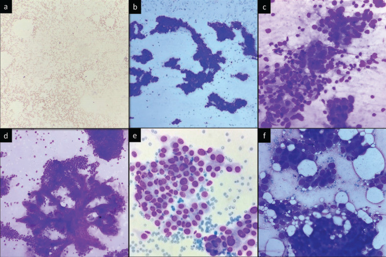
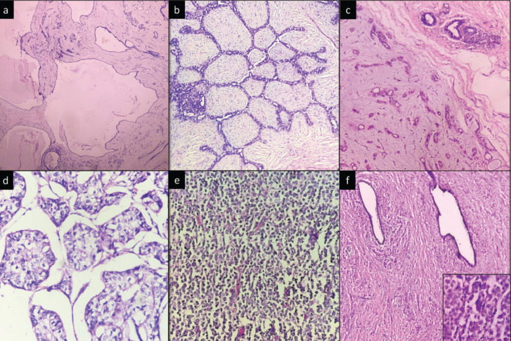
Materials and methods: A prospective study about breast FNAC was done over a period of one year, from October 2022 to September 2023. The study involved patients with breast lesion referred for FNAC in the department of Pathology. The FNAC results were further classified using the Yokohama system for reporting breast cytopathology, 2016. The cytological findings were correlated with available histopathological results.
Results: In the study of 104 cases, 60 (57.7%) of whom had available histopathology results, breast lesions were categorized using the Yokohama system as: 7.7% insufficient, 47.1% benign, 26.9% atypical, 2.9% suspicious of malignancy, and 15.4% malignant. The risk of malignancy varied by category: 0% for category 1, 3.2% for category 2, 47% for category 3, and 100% for categories 4 and 5. The maximum sensitivity was 94.7% when considering atypical, suspicious, and malignant cases as positive. The highest specificity was 97.56% for malignant cases alone, while the best diagnostic accuracy was 83.3% when both malignant and suspicious cases were counted as positive.
Conclusion: The Yokohama system effectively classified borderline lesions, facilitating early detection and improved management options. By integrating FNAC with standardized reporting, healthcare providers can make informed decisions, enhancing the diagnosis and treatment of breast lesions.

 求助内容:
求助内容: 应助结果提醒方式:


