Michael Hafner, Franziska Eckardt, Jakob Siedlecki, Benedikt Schworm, Tina R Herold, Ben Asani, Siegfried G Priglinger, Johannes B Schiefelbein
{"title":"深度学习辅助分析难治性血管性AMD改用法昔单抗后的生物标志物变化。","authors":"Michael Hafner, Franziska Eckardt, Jakob Siedlecki, Benedikt Schworm, Tina R Herold, Ben Asani, Siegfried G Priglinger, Johannes B Schiefelbein","doi":"10.1186/s40942-025-00669-2","DOIUrl":null,"url":null,"abstract":"<p><strong>Background: </strong>Artificial intelligence (AI)-driven biomarker segmentation offers an objective and reproducible approach for quantifying key anatomical features in neovascular age-related macular degeneration (nAMD) using optical coherence tomography (OCT). Currently, Faricimab, a novel bispecific inhibitor of vascular endothelial growth factor (VEGF) and angiopoietin-2 (Ang-2), offers new potential in the management of nAMD, particularly in treatment-resistant cases. This study utilizes an advanced deep learning-based segmentation algorithm to analyze OCT biomarkers and evaluate the efficacy and durability of Faricimab over nine months in patients with therapy-refractory nAMD.</p><p><strong>Methods: </strong>This retrospective real-world study analyzed patients with treatment-resistant nAMD who switched to Faricimab following inadequate responses to ranibizumab or aflibercept. Automated segmentation of key OCT biomarkers - including fibrovascular pigment epithelium detachment (fvPED), intraretinal fluid (IRF), subretinal fluid (SRF), subretinal hyperreflective material (SHRM), choroidal volume, and central retinal thickness (CRT) - was conducted using a deep learning algorithm based on a convolutional neural network.</p><p><strong>Results: </strong>A total of 46 eyes from 41 patients completed the nine-month follow-up. Significant reductions in SRF, fvPED, and choroidal volume were observed from baseline (mo0) to three months (mo3) and sustained at nine months (mo9). CRT decreased significantly from 342.7 (interquartile range (iqr): 117.1) µm at mo0 to 296.6 (iqr: 84.3) µm at mo3 and 310.2 (iqr: 93.6) µm at mo9. The deep learning model provided precise quantification of biomarkers, enabling reliable tracking of disease progression. The median injection interval extended from 35 (iqr: 15) days at mo0 to 56 (iqr: 20) days at mo9, representing a 60% increase. Visual acuity remained stable throughout the study. Correlation analysis revealed that higher baseline CRT and fvPED volumes were associated with greater best-corrected visual acuity (BCVA) improvements and longer treatment intervals.</p><p><strong>Conclusions: </strong>This study highlights the potential of AI-driven biomarker segmentation as a precise and scalable tool for monitoring disease progression in treatment-resistant nAMD. By enabling objective and reproducible analysis of OCT biomarkers, deep learning algorithms provide critical insights into treatment response. Faricimab demonstrated significant and sustained anatomical improvements, allowing for extended treatment intervals while maintaining disease stability. Future research should focus on refining AI models to improve predictive accuracy and assessing long-term outcomes to further optimize disease management.</p><p><strong>Trial registration: </strong>Ethics approval was obtained from the Institutional Review Board of LMU Munich (study ID: 20-0382). This study was conducted in accordance with the Declaration of Helsinki.</p>","PeriodicalId":14289,"journal":{"name":"International Journal of Retina and Vitreous","volume":"11 1","pages":"44"},"PeriodicalIF":2.4000,"publicationDate":"2025-04-11","publicationTypes":"Journal Article","fieldsOfStudy":null,"isOpenAccess":false,"openAccessPdf":"https://www.ncbi.nlm.nih.gov/pmc/articles/PMC11992866/pdf/","citationCount":"0","resultStr":"{\"title\":\"Deep learning assisted analysis of biomarker changes in refractory neovascular AMD after switch to faricimab.\",\"authors\":\"Michael Hafner, Franziska Eckardt, Jakob Siedlecki, Benedikt Schworm, Tina R Herold, Ben Asani, Siegfried G Priglinger, Johannes B Schiefelbein\",\"doi\":\"10.1186/s40942-025-00669-2\",\"DOIUrl\":null,\"url\":null,\"abstract\":\"<p><strong>Background: </strong>Artificial intelligence (AI)-driven biomarker segmentation offers an objective and reproducible approach for quantifying key anatomical features in neovascular age-related macular degeneration (nAMD) using optical coherence tomography (OCT). Currently, Faricimab, a novel bispecific inhibitor of vascular endothelial growth factor (VEGF) and angiopoietin-2 (Ang-2), offers new potential in the management of nAMD, particularly in treatment-resistant cases. This study utilizes an advanced deep learning-based segmentation algorithm to analyze OCT biomarkers and evaluate the efficacy and durability of Faricimab over nine months in patients with therapy-refractory nAMD.</p><p><strong>Methods: </strong>This retrospective real-world study analyzed patients with treatment-resistant nAMD who switched to Faricimab following inadequate responses to ranibizumab or aflibercept. Automated segmentation of key OCT biomarkers - including fibrovascular pigment epithelium detachment (fvPED), intraretinal fluid (IRF), subretinal fluid (SRF), subretinal hyperreflective material (SHRM), choroidal volume, and central retinal thickness (CRT) - was conducted using a deep learning algorithm based on a convolutional neural network.</p><p><strong>Results: </strong>A total of 46 eyes from 41 patients completed the nine-month follow-up. Significant reductions in SRF, fvPED, and choroidal volume were observed from baseline (mo0) to three months (mo3) and sustained at nine months (mo9). CRT decreased significantly from 342.7 (interquartile range (iqr): 117.1) µm at mo0 to 296.6 (iqr: 84.3) µm at mo3 and 310.2 (iqr: 93.6) µm at mo9. The deep learning model provided precise quantification of biomarkers, enabling reliable tracking of disease progression. The median injection interval extended from 35 (iqr: 15) days at mo0 to 56 (iqr: 20) days at mo9, representing a 60% increase. Visual acuity remained stable throughout the study. Correlation analysis revealed that higher baseline CRT and fvPED volumes were associated with greater best-corrected visual acuity (BCVA) improvements and longer treatment intervals.</p><p><strong>Conclusions: </strong>This study highlights the potential of AI-driven biomarker segmentation as a precise and scalable tool for monitoring disease progression in treatment-resistant nAMD. By enabling objective and reproducible analysis of OCT biomarkers, deep learning algorithms provide critical insights into treatment response. Faricimab demonstrated significant and sustained anatomical improvements, allowing for extended treatment intervals while maintaining disease stability. Future research should focus on refining AI models to improve predictive accuracy and assessing long-term outcomes to further optimize disease management.</p><p><strong>Trial registration: </strong>Ethics approval was obtained from the Institutional Review Board of LMU Munich (study ID: 20-0382). This study was conducted in accordance with the Declaration of Helsinki.</p>\",\"PeriodicalId\":14289,\"journal\":{\"name\":\"International Journal of Retina and Vitreous\",\"volume\":\"11 1\",\"pages\":\"44\"},\"PeriodicalIF\":2.4000,\"publicationDate\":\"2025-04-11\",\"publicationTypes\":\"Journal Article\",\"fieldsOfStudy\":null,\"isOpenAccess\":false,\"openAccessPdf\":\"https://www.ncbi.nlm.nih.gov/pmc/articles/PMC11992866/pdf/\",\"citationCount\":\"0\",\"resultStr\":null,\"platform\":\"Semanticscholar\",\"paperid\":null,\"PeriodicalName\":\"International Journal of Retina and Vitreous\",\"FirstCategoryId\":\"1085\",\"ListUrlMain\":\"https://doi.org/10.1186/s40942-025-00669-2\",\"RegionNum\":0,\"RegionCategory\":null,\"ArticlePicture\":[],\"TitleCN\":null,\"AbstractTextCN\":null,\"PMCID\":null,\"EPubDate\":\"\",\"PubModel\":\"\",\"JCR\":\"Q2\",\"JCRName\":\"OPHTHALMOLOGY\",\"Score\":null,\"Total\":0}","platform":"Semanticscholar","paperid":null,"PeriodicalName":"International Journal of Retina and Vitreous","FirstCategoryId":"1085","ListUrlMain":"https://doi.org/10.1186/s40942-025-00669-2","RegionNum":0,"RegionCategory":null,"ArticlePicture":[],"TitleCN":null,"AbstractTextCN":null,"PMCID":null,"EPubDate":"","PubModel":"","JCR":"Q2","JCRName":"OPHTHALMOLOGY","Score":null,"Total":0}
Deep learning assisted analysis of biomarker changes in refractory neovascular AMD after switch to faricimab.
Background: Artificial intelligence (AI)-driven biomarker segmentation offers an objective and reproducible approach for quantifying key anatomical features in neovascular age-related macular degeneration (nAMD) using optical coherence tomography (OCT). Currently, Faricimab, a novel bispecific inhibitor of vascular endothelial growth factor (VEGF) and angiopoietin-2 (Ang-2), offers new potential in the management of nAMD, particularly in treatment-resistant cases. This study utilizes an advanced deep learning-based segmentation algorithm to analyze OCT biomarkers and evaluate the efficacy and durability of Faricimab over nine months in patients with therapy-refractory nAMD.
Methods: This retrospective real-world study analyzed patients with treatment-resistant nAMD who switched to Faricimab following inadequate responses to ranibizumab or aflibercept. Automated segmentation of key OCT biomarkers - including fibrovascular pigment epithelium detachment (fvPED), intraretinal fluid (IRF), subretinal fluid (SRF), subretinal hyperreflective material (SHRM), choroidal volume, and central retinal thickness (CRT) - was conducted using a deep learning algorithm based on a convolutional neural network.
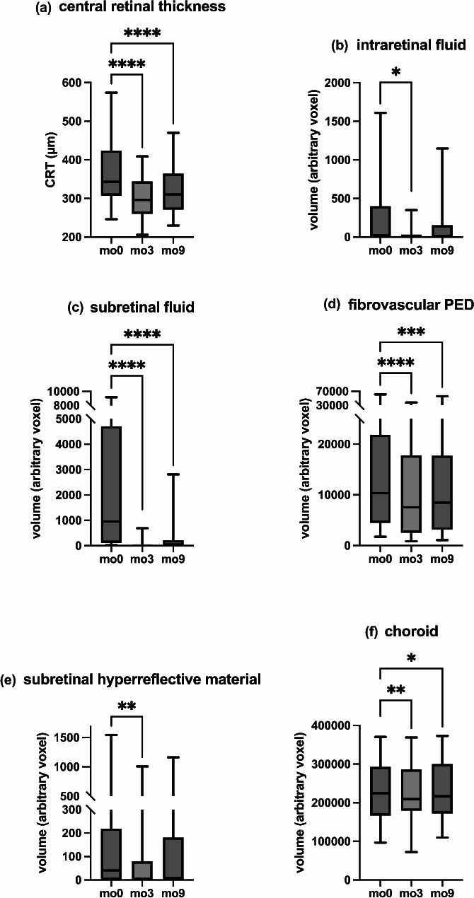
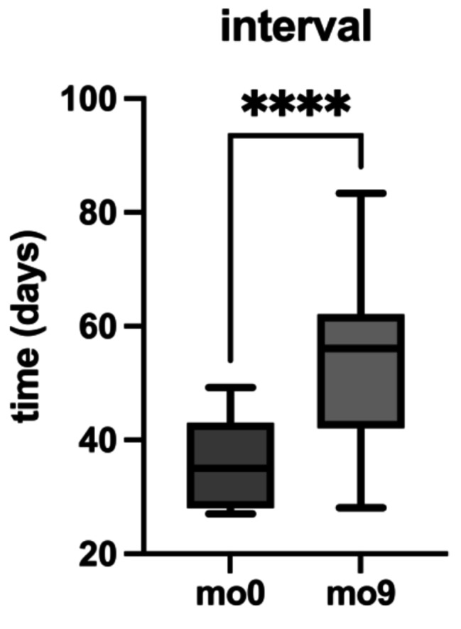
Results: A total of 46 eyes from 41 patients completed the nine-month follow-up. Significant reductions in SRF, fvPED, and choroidal volume were observed from baseline (mo0) to three months (mo3) and sustained at nine months (mo9). CRT decreased significantly from 342.7 (interquartile range (iqr): 117.1) µm at mo0 to 296.6 (iqr: 84.3) µm at mo3 and 310.2 (iqr: 93.6) µm at mo9. The deep learning model provided precise quantification of biomarkers, enabling reliable tracking of disease progression. The median injection interval extended from 35 (iqr: 15) days at mo0 to 56 (iqr: 20) days at mo9, representing a 60% increase. Visual acuity remained stable throughout the study. Correlation analysis revealed that higher baseline CRT and fvPED volumes were associated with greater best-corrected visual acuity (BCVA) improvements and longer treatment intervals.
Conclusions: This study highlights the potential of AI-driven biomarker segmentation as a precise and scalable tool for monitoring disease progression in treatment-resistant nAMD. By enabling objective and reproducible analysis of OCT biomarkers, deep learning algorithms provide critical insights into treatment response. Faricimab demonstrated significant and sustained anatomical improvements, allowing for extended treatment intervals while maintaining disease stability. Future research should focus on refining AI models to improve predictive accuracy and assessing long-term outcomes to further optimize disease management.
Trial registration: Ethics approval was obtained from the Institutional Review Board of LMU Munich (study ID: 20-0382). This study was conducted in accordance with the Declaration of Helsinki.
期刊介绍:
International Journal of Retina and Vitreous focuses on the ophthalmic subspecialty of vitreoretinal disorders. The journal presents original articles on new approaches to diagnosis, outcomes of clinical trials, innovations in pharmacological therapy and surgical techniques, as well as basic science advances that impact clinical practice. Topical areas include, but are not limited to: -Imaging of the retina, choroid and vitreous -Innovations in optical coherence tomography (OCT) -Small-gauge vitrectomy, retinal detachment, chromovitrectomy -Electroretinography (ERG), microperimetry, other functional tests -Intraocular tumors -Retinal pharmacotherapy & drug delivery -Diabetic retinopathy & other vascular diseases -Age-related macular degeneration (AMD) & other macular entities

 求助内容:
求助内容: 应助结果提醒方式:


