{"title":"von Hippel-Lindau病视网膜血管增生伴纤维化消退。","authors":"Jing-Yi Chen, Hung-Da Chou, An-Ning Chao, Chi-Chun Lai, Mandeep S Sagoo","doi":"10.4103/tjo.TJO-D-24-00116","DOIUrl":null,"url":null,"abstract":"<p><p>In this study, we report a rare case of retinal vascular proliferation (RVP) in von Hippel-Lindau (VHL) disease, followed by a literature review. A 12-year-old boy presented with a left cerebellar hemangioblastoma and right eye blurred vision for 1-2 years. Fundus examination found no capillary hemangioblastoma lesion but a broad epiretinal fibrovascular membrane, which caused significant traction to the right macula. The genetic testing identified a pathogenic missense mutation (c. 223A > G) within the <i>VHL</i> gene, confirming VHL disease. RVP is a less common, poorly understood condition that can occur in VHL disease apart from the typical retinal capillary hemangioblastoma. The surface vasculature of the fibrovascular membrane regressed over an observation period of 3 years, and pars plana vitrectomy was eventually conducted at the age of 15 years to remove the fibrovascular membrane. Nevertheless, his visual acuity remained at 20/200 at postoperative 1 year due to the development of cataracts. In our literature review, we analyzed 39 reported cases of RVP, of which 90% had unilateral lesions, 70% had lesions at the juxtapapillary location, and 50% had a visual acuity <20/40. The mean onset age was 24 years. An intervention was performed in 39% of the cases and 78% experienced improved vision posttreatment. In conclusion, RVP likely starts as mainly vascular proliferation and eventually regresses spontaneously to fibrotic tissue formation. Unlike typical retinal capillary hemangioblastoma, vision can improve after an intervention, even in eyes with juxtapapillary lesions.</p>","PeriodicalId":44978,"journal":{"name":"Taiwan Journal of Ophthalmology","volume":"15 1","pages":"138-142"},"PeriodicalIF":1.2000,"publicationDate":"2025-03-05","publicationTypes":"Journal Article","fieldsOfStudy":null,"isOpenAccess":false,"openAccessPdf":"https://www.ncbi.nlm.nih.gov/pmc/articles/PMC11981565/pdf/","citationCount":"0","resultStr":"{\"title\":\"Retinal vascular proliferation with fibrotic regression in von Hippel-Lindau disease.\",\"authors\":\"Jing-Yi Chen, Hung-Da Chou, An-Ning Chao, Chi-Chun Lai, Mandeep S Sagoo\",\"doi\":\"10.4103/tjo.TJO-D-24-00116\",\"DOIUrl\":null,\"url\":null,\"abstract\":\"<p><p>In this study, we report a rare case of retinal vascular proliferation (RVP) in von Hippel-Lindau (VHL) disease, followed by a literature review. A 12-year-old boy presented with a left cerebellar hemangioblastoma and right eye blurred vision for 1-2 years. Fundus examination found no capillary hemangioblastoma lesion but a broad epiretinal fibrovascular membrane, which caused significant traction to the right macula. The genetic testing identified a pathogenic missense mutation (c. 223A > G) within the <i>VHL</i> gene, confirming VHL disease. RVP is a less common, poorly understood condition that can occur in VHL disease apart from the typical retinal capillary hemangioblastoma. The surface vasculature of the fibrovascular membrane regressed over an observation period of 3 years, and pars plana vitrectomy was eventually conducted at the age of 15 years to remove the fibrovascular membrane. Nevertheless, his visual acuity remained at 20/200 at postoperative 1 year due to the development of cataracts. In our literature review, we analyzed 39 reported cases of RVP, of which 90% had unilateral lesions, 70% had lesions at the juxtapapillary location, and 50% had a visual acuity <20/40. The mean onset age was 24 years. An intervention was performed in 39% of the cases and 78% experienced improved vision posttreatment. In conclusion, RVP likely starts as mainly vascular proliferation and eventually regresses spontaneously to fibrotic tissue formation. Unlike typical retinal capillary hemangioblastoma, vision can improve after an intervention, even in eyes with juxtapapillary lesions.</p>\",\"PeriodicalId\":44978,\"journal\":{\"name\":\"Taiwan Journal of Ophthalmology\",\"volume\":\"15 1\",\"pages\":\"138-142\"},\"PeriodicalIF\":1.2000,\"publicationDate\":\"2025-03-05\",\"publicationTypes\":\"Journal Article\",\"fieldsOfStudy\":null,\"isOpenAccess\":false,\"openAccessPdf\":\"https://www.ncbi.nlm.nih.gov/pmc/articles/PMC11981565/pdf/\",\"citationCount\":\"0\",\"resultStr\":null,\"platform\":\"Semanticscholar\",\"paperid\":null,\"PeriodicalName\":\"Taiwan Journal of Ophthalmology\",\"FirstCategoryId\":\"1085\",\"ListUrlMain\":\"https://doi.org/10.4103/tjo.TJO-D-24-00116\",\"RegionNum\":0,\"RegionCategory\":null,\"ArticlePicture\":[],\"TitleCN\":null,\"AbstractTextCN\":null,\"PMCID\":null,\"EPubDate\":\"2025/1/1 0:00:00\",\"PubModel\":\"eCollection\",\"JCR\":\"Q4\",\"JCRName\":\"OPHTHALMOLOGY\",\"Score\":null,\"Total\":0}","platform":"Semanticscholar","paperid":null,"PeriodicalName":"Taiwan Journal of Ophthalmology","FirstCategoryId":"1085","ListUrlMain":"https://doi.org/10.4103/tjo.TJO-D-24-00116","RegionNum":0,"RegionCategory":null,"ArticlePicture":[],"TitleCN":null,"AbstractTextCN":null,"PMCID":null,"EPubDate":"2025/1/1 0:00:00","PubModel":"eCollection","JCR":"Q4","JCRName":"OPHTHALMOLOGY","Score":null,"Total":0}
Retinal vascular proliferation with fibrotic regression in von Hippel-Lindau disease.
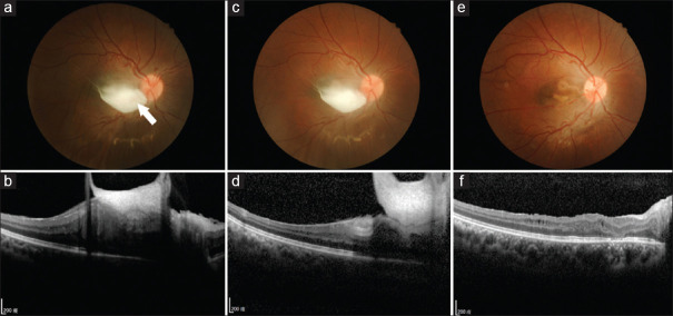
In this study, we report a rare case of retinal vascular proliferation (RVP) in von Hippel-Lindau (VHL) disease, followed by a literature review. A 12-year-old boy presented with a left cerebellar hemangioblastoma and right eye blurred vision for 1-2 years. Fundus examination found no capillary hemangioblastoma lesion but a broad epiretinal fibrovascular membrane, which caused significant traction to the right macula. The genetic testing identified a pathogenic missense mutation (c. 223A > G) within the VHL gene, confirming VHL disease. RVP is a less common, poorly understood condition that can occur in VHL disease apart from the typical retinal capillary hemangioblastoma. The surface vasculature of the fibrovascular membrane regressed over an observation period of 3 years, and pars plana vitrectomy was eventually conducted at the age of 15 years to remove the fibrovascular membrane. Nevertheless, his visual acuity remained at 20/200 at postoperative 1 year due to the development of cataracts. In our literature review, we analyzed 39 reported cases of RVP, of which 90% had unilateral lesions, 70% had lesions at the juxtapapillary location, and 50% had a visual acuity <20/40. The mean onset age was 24 years. An intervention was performed in 39% of the cases and 78% experienced improved vision posttreatment. In conclusion, RVP likely starts as mainly vascular proliferation and eventually regresses spontaneously to fibrotic tissue formation. Unlike typical retinal capillary hemangioblastoma, vision can improve after an intervention, even in eyes with juxtapapillary lesions.

 求助内容:
求助内容: 应助结果提醒方式:


