Salim S Alkeraye, Turky N Alsehli, Ahmed Alhumidi, Khalid Al-Husain, Khalid Nabil Nagshabandi
{"title":"罕见蕈样真菌病伴角化孔样病变1例报告及文献复习。","authors":"Salim S Alkeraye, Turky N Alsehli, Ahmed Alhumidi, Khalid Al-Husain, Khalid Nabil Nagshabandi","doi":"10.21037/acr-24-210","DOIUrl":null,"url":null,"abstract":"<p><strong>Background: </strong>Cutaneous T-cell lymphomas (CTCLs) are the second most prevalent group of extranodal lymphomas second to B-cell lymphomas. Mycosis fungoides (MF) is the most common type of CTCL, often presenting as erythematous patches and plaques with scaling. However, MF is known for its wide range of clinical presentations, making it a \"great mimicker\" of other dermatological conditions. It can manifest as hypopigmented, hyperkeratotic, or purpuric lesions, among others, contributing to frequent diagnostic challenges. Porokeratosis-like MF is an exceedingly rare variant, with only a few cases reported in the literature. This case report describes a unique presentation of MF mimicking porokeratosis.</p><p><strong>Case description: </strong>A 44-year-old male presented with brownish papules on the feet, showing porokeratosis-like features upon clinical presentation. Histopathological examination and immunohistochemistry revealed a profile consistent with MF, demonstrating band-like infiltrate of lymphocytes in the papillary dermis and CD7 loss and CD8 positivity in both the epidermis and dermis. The patient was treated with topical Clobetasol propionate, showing partial improvement over a biweekly follow-up period of three months with no recurrence observed to date.</p><p><strong>Conclusions: </strong>This case underscores the importance of considering MF in atypical porokeratosis presentations. A thorough clinicopathologic correlation is vital for accurate diagnosis and management of such unusual MF variants.</p>","PeriodicalId":29752,"journal":{"name":"AME Case Reports","volume":"9 ","pages":"67"},"PeriodicalIF":0.7000,"publicationDate":"2025-03-31","publicationTypes":"Journal Article","fieldsOfStudy":null,"isOpenAccess":false,"openAccessPdf":"https://www.ncbi.nlm.nih.gov/pmc/articles/PMC12053989/pdf/","citationCount":"0","resultStr":"{\"title\":\"A rare case of mycosis fungoides with porokeratosis-like lesions: a case report and review of previous literature.\",\"authors\":\"Salim S Alkeraye, Turky N Alsehli, Ahmed Alhumidi, Khalid Al-Husain, Khalid Nabil Nagshabandi\",\"doi\":\"10.21037/acr-24-210\",\"DOIUrl\":null,\"url\":null,\"abstract\":\"<p><strong>Background: </strong>Cutaneous T-cell lymphomas (CTCLs) are the second most prevalent group of extranodal lymphomas second to B-cell lymphomas. Mycosis fungoides (MF) is the most common type of CTCL, often presenting as erythematous patches and plaques with scaling. However, MF is known for its wide range of clinical presentations, making it a \\\"great mimicker\\\" of other dermatological conditions. It can manifest as hypopigmented, hyperkeratotic, or purpuric lesions, among others, contributing to frequent diagnostic challenges. Porokeratosis-like MF is an exceedingly rare variant, with only a few cases reported in the literature. This case report describes a unique presentation of MF mimicking porokeratosis.</p><p><strong>Case description: </strong>A 44-year-old male presented with brownish papules on the feet, showing porokeratosis-like features upon clinical presentation. Histopathological examination and immunohistochemistry revealed a profile consistent with MF, demonstrating band-like infiltrate of lymphocytes in the papillary dermis and CD7 loss and CD8 positivity in both the epidermis and dermis. The patient was treated with topical Clobetasol propionate, showing partial improvement over a biweekly follow-up period of three months with no recurrence observed to date.</p><p><strong>Conclusions: </strong>This case underscores the importance of considering MF in atypical porokeratosis presentations. A thorough clinicopathologic correlation is vital for accurate diagnosis and management of such unusual MF variants.</p>\",\"PeriodicalId\":29752,\"journal\":{\"name\":\"AME Case Reports\",\"volume\":\"9 \",\"pages\":\"67\"},\"PeriodicalIF\":0.7000,\"publicationDate\":\"2025-03-31\",\"publicationTypes\":\"Journal Article\",\"fieldsOfStudy\":null,\"isOpenAccess\":false,\"openAccessPdf\":\"https://www.ncbi.nlm.nih.gov/pmc/articles/PMC12053989/pdf/\",\"citationCount\":\"0\",\"resultStr\":null,\"platform\":\"Semanticscholar\",\"paperid\":null,\"PeriodicalName\":\"AME Case Reports\",\"FirstCategoryId\":\"1085\",\"ListUrlMain\":\"https://doi.org/10.21037/acr-24-210\",\"RegionNum\":0,\"RegionCategory\":null,\"ArticlePicture\":[],\"TitleCN\":null,\"AbstractTextCN\":null,\"PMCID\":null,\"EPubDate\":\"2025/1/1 0:00:00\",\"PubModel\":\"eCollection\",\"JCR\":\"Q3\",\"JCRName\":\"MEDICINE, GENERAL & INTERNAL\",\"Score\":null,\"Total\":0}","platform":"Semanticscholar","paperid":null,"PeriodicalName":"AME Case Reports","FirstCategoryId":"1085","ListUrlMain":"https://doi.org/10.21037/acr-24-210","RegionNum":0,"RegionCategory":null,"ArticlePicture":[],"TitleCN":null,"AbstractTextCN":null,"PMCID":null,"EPubDate":"2025/1/1 0:00:00","PubModel":"eCollection","JCR":"Q3","JCRName":"MEDICINE, GENERAL & INTERNAL","Score":null,"Total":0}
A rare case of mycosis fungoides with porokeratosis-like lesions: a case report and review of previous literature.
Background: Cutaneous T-cell lymphomas (CTCLs) are the second most prevalent group of extranodal lymphomas second to B-cell lymphomas. Mycosis fungoides (MF) is the most common type of CTCL, often presenting as erythematous patches and plaques with scaling. However, MF is known for its wide range of clinical presentations, making it a "great mimicker" of other dermatological conditions. It can manifest as hypopigmented, hyperkeratotic, or purpuric lesions, among others, contributing to frequent diagnostic challenges. Porokeratosis-like MF is an exceedingly rare variant, with only a few cases reported in the literature. This case report describes a unique presentation of MF mimicking porokeratosis.
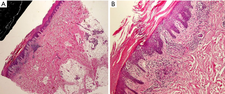
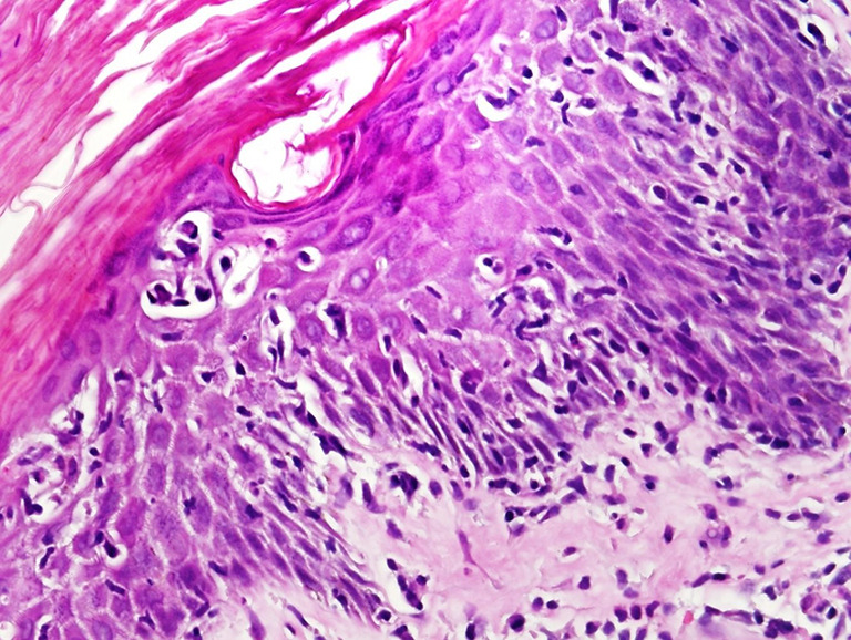
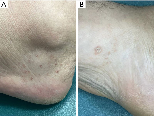
Case description: A 44-year-old male presented with brownish papules on the feet, showing porokeratosis-like features upon clinical presentation. Histopathological examination and immunohistochemistry revealed a profile consistent with MF, demonstrating band-like infiltrate of lymphocytes in the papillary dermis and CD7 loss and CD8 positivity in both the epidermis and dermis. The patient was treated with topical Clobetasol propionate, showing partial improvement over a biweekly follow-up period of three months with no recurrence observed to date.
Conclusions: This case underscores the importance of considering MF in atypical porokeratosis presentations. A thorough clinicopathologic correlation is vital for accurate diagnosis and management of such unusual MF variants.

 求助内容:
求助内容: 应助结果提醒方式:


