Duygu Ozgul, Ferhat Can Piskin, Sinan Sozutok, Huseyin Tugsan Balli
{"title":"肝细胞癌患者肝功能影像学评分的验证:加多西酸增强MRI。","authors":"Duygu Ozgul, Ferhat Can Piskin, Sinan Sozutok, Huseyin Tugsan Balli","doi":"10.14744/hf.2023.2023.0024","DOIUrl":null,"url":null,"abstract":"<p><strong>Background and aim: </strong>To investigate the correlation of the functional liver imaging scores (FLIS) and the scoring system in hepatocellular carcinoma (HCC) patients.</p><p><strong>Materials and methods: </strong>Between April 2015 and December 2022, the HCC patients who underwent gadoxetic acid-enhanced MRI were analyzed. Three parameters on hepatobiliary phase images were evaluated for FLIS: liver parenchymal enhancement, biliary excretion, and signal intensity of the portal vein. The correlation between Child-Turcotte-Pugh (CTP) classification, the albumin-bilirubin (ALBI) grade, and Fibrosis-4 (F-4) score, and FLIS were analyzed. Receiver operating characteristic curve analysis was performed to demonstrate the cut-off value of FLIS for differentiating between CTP classification and ALBI grade.</p><p><strong>Results: </strong>We retrospectively analyzed 178 HCC patients (144 men, 34 women; mean age, 65.9 years). A moderate negative correlation was present between CTP classification and ALBI grade, and FLIS (r=-0.596 and r=-0.513, respectively). FLIS ≤3 was determined as the most optimal criterion for differentiating CTP A or B patients from CTP C patients.</p><p><strong>Conclusion: </strong>This study showed that the FLIS is a simple, non-invasive imaging marker for the assessment of liver function in HCC patients.</p>","PeriodicalId":29722,"journal":{"name":"Hepatology Forum","volume":"6 2","pages":"41-46"},"PeriodicalIF":2.1000,"publicationDate":"2024-10-11","publicationTypes":"Journal Article","fieldsOfStudy":null,"isOpenAccess":false,"openAccessPdf":"https://www.ncbi.nlm.nih.gov/pmc/articles/PMC11999903/pdf/","citationCount":"0","resultStr":"{\"title\":\"Validation of functional liver imaging scores derived on gadoxetic acid-enhanced MRI in hepatocellular carcinoma patients.\",\"authors\":\"Duygu Ozgul, Ferhat Can Piskin, Sinan Sozutok, Huseyin Tugsan Balli\",\"doi\":\"10.14744/hf.2023.2023.0024\",\"DOIUrl\":null,\"url\":null,\"abstract\":\"<p><strong>Background and aim: </strong>To investigate the correlation of the functional liver imaging scores (FLIS) and the scoring system in hepatocellular carcinoma (HCC) patients.</p><p><strong>Materials and methods: </strong>Between April 2015 and December 2022, the HCC patients who underwent gadoxetic acid-enhanced MRI were analyzed. Three parameters on hepatobiliary phase images were evaluated for FLIS: liver parenchymal enhancement, biliary excretion, and signal intensity of the portal vein. The correlation between Child-Turcotte-Pugh (CTP) classification, the albumin-bilirubin (ALBI) grade, and Fibrosis-4 (F-4) score, and FLIS were analyzed. Receiver operating characteristic curve analysis was performed to demonstrate the cut-off value of FLIS for differentiating between CTP classification and ALBI grade.</p><p><strong>Results: </strong>We retrospectively analyzed 178 HCC patients (144 men, 34 women; mean age, 65.9 years). A moderate negative correlation was present between CTP classification and ALBI grade, and FLIS (r=-0.596 and r=-0.513, respectively). FLIS ≤3 was determined as the most optimal criterion for differentiating CTP A or B patients from CTP C patients.</p><p><strong>Conclusion: </strong>This study showed that the FLIS is a simple, non-invasive imaging marker for the assessment of liver function in HCC patients.</p>\",\"PeriodicalId\":29722,\"journal\":{\"name\":\"Hepatology Forum\",\"volume\":\"6 2\",\"pages\":\"41-46\"},\"PeriodicalIF\":2.1000,\"publicationDate\":\"2024-10-11\",\"publicationTypes\":\"Journal Article\",\"fieldsOfStudy\":null,\"isOpenAccess\":false,\"openAccessPdf\":\"https://www.ncbi.nlm.nih.gov/pmc/articles/PMC11999903/pdf/\",\"citationCount\":\"0\",\"resultStr\":null,\"platform\":\"Semanticscholar\",\"paperid\":null,\"PeriodicalName\":\"Hepatology Forum\",\"FirstCategoryId\":\"1085\",\"ListUrlMain\":\"https://doi.org/10.14744/hf.2023.2023.0024\",\"RegionNum\":0,\"RegionCategory\":null,\"ArticlePicture\":[],\"TitleCN\":null,\"AbstractTextCN\":null,\"PMCID\":null,\"EPubDate\":\"2025/1/1 0:00:00\",\"PubModel\":\"eCollection\",\"JCR\":\"Q4\",\"JCRName\":\"GASTROENTEROLOGY & HEPATOLOGY\",\"Score\":null,\"Total\":0}","platform":"Semanticscholar","paperid":null,"PeriodicalName":"Hepatology Forum","FirstCategoryId":"1085","ListUrlMain":"https://doi.org/10.14744/hf.2023.2023.0024","RegionNum":0,"RegionCategory":null,"ArticlePicture":[],"TitleCN":null,"AbstractTextCN":null,"PMCID":null,"EPubDate":"2025/1/1 0:00:00","PubModel":"eCollection","JCR":"Q4","JCRName":"GASTROENTEROLOGY & HEPATOLOGY","Score":null,"Total":0}
Validation of functional liver imaging scores derived on gadoxetic acid-enhanced MRI in hepatocellular carcinoma patients.
Background and aim: To investigate the correlation of the functional liver imaging scores (FLIS) and the scoring system in hepatocellular carcinoma (HCC) patients.
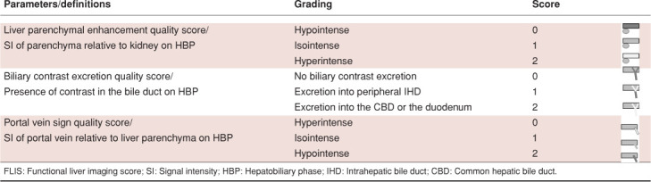
Materials and methods: Between April 2015 and December 2022, the HCC patients who underwent gadoxetic acid-enhanced MRI were analyzed. Three parameters on hepatobiliary phase images were evaluated for FLIS: liver parenchymal enhancement, biliary excretion, and signal intensity of the portal vein. The correlation between Child-Turcotte-Pugh (CTP) classification, the albumin-bilirubin (ALBI) grade, and Fibrosis-4 (F-4) score, and FLIS were analyzed. Receiver operating characteristic curve analysis was performed to demonstrate the cut-off value of FLIS for differentiating between CTP classification and ALBI grade.
Results: We retrospectively analyzed 178 HCC patients (144 men, 34 women; mean age, 65.9 years). A moderate negative correlation was present between CTP classification and ALBI grade, and FLIS (r=-0.596 and r=-0.513, respectively). FLIS ≤3 was determined as the most optimal criterion for differentiating CTP A or B patients from CTP C patients.
Conclusion: This study showed that the FLIS is a simple, non-invasive imaging marker for the assessment of liver function in HCC patients.

 求助内容:
求助内容: 应助结果提醒方式:


