Lu Qiu, Miaoyan Wang, Surui Liu, Bo Peng, Ying Hua, Jianbiao Wang, Xiaoyue Hu, Anqi Qiu, Yakang Dai, Haoxiang Jiang
{"title":"多参数MRI评价儿童脑淋巴功能障碍和脑白质异常及鉴别难治性癫痫。","authors":"Lu Qiu, Miaoyan Wang, Surui Liu, Bo Peng, Ying Hua, Jianbiao Wang, Xiaoyue Hu, Anqi Qiu, Yakang Dai, Haoxiang Jiang","doi":"10.3348/kjr.2024.0718","DOIUrl":null,"url":null,"abstract":"<p><strong>Objective: </strong>To explore glymphatic impairment in pediatric refractory epilepsy (RE) using multi-parameter magnetic resonance imaging (MRI), assess its relationship with white-matter (WM) abnormalities and clinical indicators, and preliminarily evaluate the performance of multi-parameter MRI in discriminating RE from drug-sensitive epilepsy (DSE).</p><p><strong>Materials and methods: </strong>We retrospectively included 70 patients with DSE (mean age, 9.7 ± 3.5 years; male:female, 37:33) and 26 patients with RE (9.0 ± 2.9 years; male:female, 12:14). The diffusion tensor imaging analysis along the perivascular space (DTI-ALPS) index as well as fractional anisotropy (FA), mean diffusivity (MD), and nodal efficiency values were measured and compared between patients with RE and DSE. With sex and age as covariables, differences in the FA and MD values were analyzed using tract-based spatial statistics, and nodal efficiency was analyzed using a linear model. Pearson's partial correlation was analyzed. Receiver operating characteristic (ROC) curves were used to evaluate the discrimination performance of the MRI-based machine-learning models through five-fold cross-validation.</p><p><strong>Results: </strong>In the RE group, FA decreased and MD increased in comparison with the corresponding values in the DSE group, and these differences mainly involved the callosum, right and left corona radiata, inferior and superior longitudinal fasciculus, and posterior thalamic radiation (threshold-free cluster enhancement, <i>P</i> < 0.05). The RE group also showed reduced nodal efficiency, which mainly involved the limbic system, default mode network, and visual network (false discovery rate, <i>P</i> < 0.05), and significantly lower DTI-ALPS index (F = 2.0, <i>P</i> = 0.049). The DTI-ALPS index was positively correlated with FA (0.25 ≤ <i>r</i> ≤ 0.32) and nodal efficiency (0.22 ≤ <i>r</i> ≤ 0.37), and was negatively correlated with the MD (-0.24 ≤ <i>r</i> ≤ -0.34) and seizure frequency (<i>r</i> = -0.47). A machine-learning model combining DTI-ALPS, FA, MD, and nodal efficiency achieved a cross-validated ROC curve area of 0.83 (sensitivity, 78.2%; specificity, 84.8%).</p><p><strong>Conclusion: </strong>Pediatric patients with RE showed impaired glymphatic function in comparison with patients with DSE, which was correlated with WM abnormalities and seizure frequency. Multi-parameter MRI may be feasible for distinguishing RE from DSE.</p>","PeriodicalId":17881,"journal":{"name":"Korean Journal of Radiology","volume":"26 5","pages":"485-497"},"PeriodicalIF":5.3000,"publicationDate":"2025-05-01","publicationTypes":"Journal Article","fieldsOfStudy":null,"isOpenAccess":false,"openAccessPdf":"https://www.ncbi.nlm.nih.gov/pmc/articles/PMC12055269/pdf/","citationCount":"0","resultStr":"{\"title\":\"Multi-Parameter MRI for Evaluating Glymphatic Impairment and White-Matter Abnormalities and Discriminating Refractory Epilepsy in Children.\",\"authors\":\"Lu Qiu, Miaoyan Wang, Surui Liu, Bo Peng, Ying Hua, Jianbiao Wang, Xiaoyue Hu, Anqi Qiu, Yakang Dai, Haoxiang Jiang\",\"doi\":\"10.3348/kjr.2024.0718\",\"DOIUrl\":null,\"url\":null,\"abstract\":\"<p><strong>Objective: </strong>To explore glymphatic impairment in pediatric refractory epilepsy (RE) using multi-parameter magnetic resonance imaging (MRI), assess its relationship with white-matter (WM) abnormalities and clinical indicators, and preliminarily evaluate the performance of multi-parameter MRI in discriminating RE from drug-sensitive epilepsy (DSE).</p><p><strong>Materials and methods: </strong>We retrospectively included 70 patients with DSE (mean age, 9.7 ± 3.5 years; male:female, 37:33) and 26 patients with RE (9.0 ± 2.9 years; male:female, 12:14). The diffusion tensor imaging analysis along the perivascular space (DTI-ALPS) index as well as fractional anisotropy (FA), mean diffusivity (MD), and nodal efficiency values were measured and compared between patients with RE and DSE. With sex and age as covariables, differences in the FA and MD values were analyzed using tract-based spatial statistics, and nodal efficiency was analyzed using a linear model. Pearson's partial correlation was analyzed. Receiver operating characteristic (ROC) curves were used to evaluate the discrimination performance of the MRI-based machine-learning models through five-fold cross-validation.</p><p><strong>Results: </strong>In the RE group, FA decreased and MD increased in comparison with the corresponding values in the DSE group, and these differences mainly involved the callosum, right and left corona radiata, inferior and superior longitudinal fasciculus, and posterior thalamic radiation (threshold-free cluster enhancement, <i>P</i> < 0.05). The RE group also showed reduced nodal efficiency, which mainly involved the limbic system, default mode network, and visual network (false discovery rate, <i>P</i> < 0.05), and significantly lower DTI-ALPS index (F = 2.0, <i>P</i> = 0.049). The DTI-ALPS index was positively correlated with FA (0.25 ≤ <i>r</i> ≤ 0.32) and nodal efficiency (0.22 ≤ <i>r</i> ≤ 0.37), and was negatively correlated with the MD (-0.24 ≤ <i>r</i> ≤ -0.34) and seizure frequency (<i>r</i> = -0.47). A machine-learning model combining DTI-ALPS, FA, MD, and nodal efficiency achieved a cross-validated ROC curve area of 0.83 (sensitivity, 78.2%; specificity, 84.8%).</p><p><strong>Conclusion: </strong>Pediatric patients with RE showed impaired glymphatic function in comparison with patients with DSE, which was correlated with WM abnormalities and seizure frequency. Multi-parameter MRI may be feasible for distinguishing RE from DSE.</p>\",\"PeriodicalId\":17881,\"journal\":{\"name\":\"Korean Journal of Radiology\",\"volume\":\"26 5\",\"pages\":\"485-497\"},\"PeriodicalIF\":5.3000,\"publicationDate\":\"2025-05-01\",\"publicationTypes\":\"Journal Article\",\"fieldsOfStudy\":null,\"isOpenAccess\":false,\"openAccessPdf\":\"https://www.ncbi.nlm.nih.gov/pmc/articles/PMC12055269/pdf/\",\"citationCount\":\"0\",\"resultStr\":null,\"platform\":\"Semanticscholar\",\"paperid\":null,\"PeriodicalName\":\"Korean Journal of Radiology\",\"FirstCategoryId\":\"3\",\"ListUrlMain\":\"https://doi.org/10.3348/kjr.2024.0718\",\"RegionNum\":2,\"RegionCategory\":\"医学\",\"ArticlePicture\":[],\"TitleCN\":null,\"AbstractTextCN\":null,\"PMCID\":null,\"EPubDate\":\"\",\"PubModel\":\"\",\"JCR\":\"Q1\",\"JCRName\":\"RADIOLOGY, NUCLEAR MEDICINE & MEDICAL IMAGING\",\"Score\":null,\"Total\":0}","platform":"Semanticscholar","paperid":null,"PeriodicalName":"Korean Journal of Radiology","FirstCategoryId":"3","ListUrlMain":"https://doi.org/10.3348/kjr.2024.0718","RegionNum":2,"RegionCategory":"医学","ArticlePicture":[],"TitleCN":null,"AbstractTextCN":null,"PMCID":null,"EPubDate":"","PubModel":"","JCR":"Q1","JCRName":"RADIOLOGY, NUCLEAR MEDICINE & MEDICAL IMAGING","Score":null,"Total":0}
Multi-Parameter MRI for Evaluating Glymphatic Impairment and White-Matter Abnormalities and Discriminating Refractory Epilepsy in Children.
Objective: To explore glymphatic impairment in pediatric refractory epilepsy (RE) using multi-parameter magnetic resonance imaging (MRI), assess its relationship with white-matter (WM) abnormalities and clinical indicators, and preliminarily evaluate the performance of multi-parameter MRI in discriminating RE from drug-sensitive epilepsy (DSE).
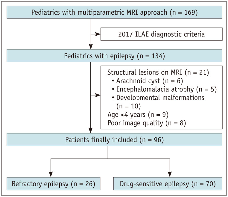
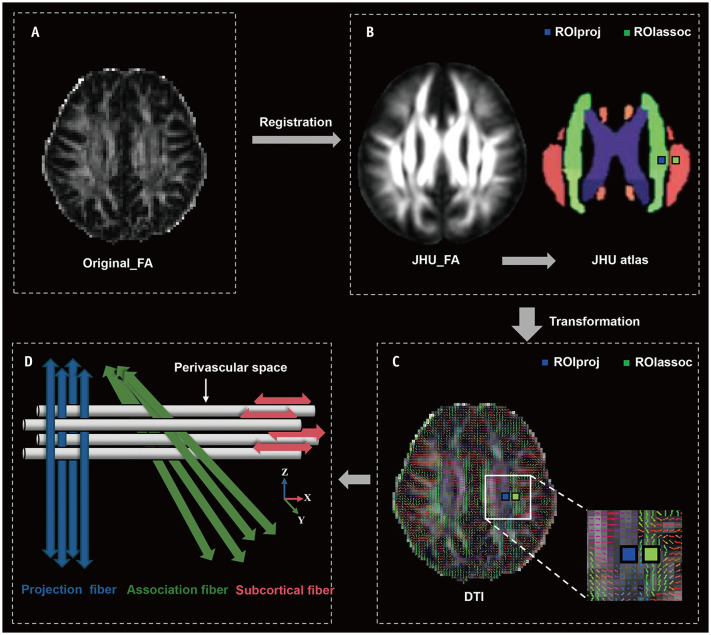
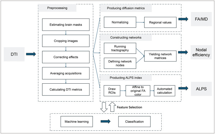
Materials and methods: We retrospectively included 70 patients with DSE (mean age, 9.7 ± 3.5 years; male:female, 37:33) and 26 patients with RE (9.0 ± 2.9 years; male:female, 12:14). The diffusion tensor imaging analysis along the perivascular space (DTI-ALPS) index as well as fractional anisotropy (FA), mean diffusivity (MD), and nodal efficiency values were measured and compared between patients with RE and DSE. With sex and age as covariables, differences in the FA and MD values were analyzed using tract-based spatial statistics, and nodal efficiency was analyzed using a linear model. Pearson's partial correlation was analyzed. Receiver operating characteristic (ROC) curves were used to evaluate the discrimination performance of the MRI-based machine-learning models through five-fold cross-validation.
Results: In the RE group, FA decreased and MD increased in comparison with the corresponding values in the DSE group, and these differences mainly involved the callosum, right and left corona radiata, inferior and superior longitudinal fasciculus, and posterior thalamic radiation (threshold-free cluster enhancement, P < 0.05). The RE group also showed reduced nodal efficiency, which mainly involved the limbic system, default mode network, and visual network (false discovery rate, P < 0.05), and significantly lower DTI-ALPS index (F = 2.0, P = 0.049). The DTI-ALPS index was positively correlated with FA (0.25 ≤ r ≤ 0.32) and nodal efficiency (0.22 ≤ r ≤ 0.37), and was negatively correlated with the MD (-0.24 ≤ r ≤ -0.34) and seizure frequency (r = -0.47). A machine-learning model combining DTI-ALPS, FA, MD, and nodal efficiency achieved a cross-validated ROC curve area of 0.83 (sensitivity, 78.2%; specificity, 84.8%).
Conclusion: Pediatric patients with RE showed impaired glymphatic function in comparison with patients with DSE, which was correlated with WM abnormalities and seizure frequency. Multi-parameter MRI may be feasible for distinguishing RE from DSE.
期刊介绍:
The inaugural issue of the Korean J Radiol came out in March 2000. Our journal aims to produce and propagate knowledge on radiologic imaging and related sciences.
A unique feature of the articles published in the Journal will be their reflection of global trends in radiology combined with an East-Asian perspective. Geographic differences in disease prevalence will be reflected in the contents of papers, and this will serve to enrich our body of knowledge.
World''s outstanding radiologists from many countries are serving as editorial board of our journal.

 求助内容:
求助内容: 应助结果提醒方式:


