Tormund H Njølstad, Kristin Jensen, Hilde K Andersen, Audun E Berstad, Gaute Hagen, Cathrine K Johansen, Kjetil Øye, Jan Glittum, Anniken Dybwad, Emma Thingstad, Marianne G Guren, Johann Baptist Dormagen, Anselm Schulz
{"title":"深度学习重建在标准剂量和减剂量腹部CT肝脏病变检测中的应用。","authors":"Tormund H Njølstad, Kristin Jensen, Hilde K Andersen, Audun E Berstad, Gaute Hagen, Cathrine K Johansen, Kjetil Øye, Jan Glittum, Anniken Dybwad, Emma Thingstad, Marianne G Guren, Johann Baptist Dormagen, Anselm Schulz","doi":"10.1007/s00330-025-11596-z","DOIUrl":null,"url":null,"abstract":"<p><strong>Objectives: </strong>Deep learning reconstruction (DLR) has shown promising image denoising ability, but its radiation dose reduction potential remains unknown. The objective of this study was to investigate the diagnostic performance of DLR compared to iterative reconstruction (IR) in the detection of liver lesions at standard-dose and reduced-dose CT.</p><p><strong>Materials and methods: </strong>Participants with known liver metastases from gastrointestinal and pancreatic adenocarcinoma were prospectively included from routine follow-up (October 2020 to March 2022). Participants received standard-dose CT and two additional reduced-dose scans during the same contrast administration, each reconstructed with IR and high-strength DLR. Two radiologists evaluated images for the presence of liver lesions, and a third established a reference standard. Diagnostic performance was compared using McNemar's test and mixed effects logistic regression.</p><p><strong>Results: </strong>Forty-four participants (mean age 66 years ± 11 [standard deviation], 28 men) were evaluated with 348 included liver lesions ≤ 20 mm (297 metastases, 51 benign; mean size 9.1 ± 4.3 mm). Mean volume CT dose index was 14.2, 7.8 mGy, and 5.1 mGy. Between algorithms, no significant difference in lesion detection was observed within dose levels. Detection of 233 lesions ≤ 10 mm was deteriorated with lower dose levels despite DLR denoising, with 185 detected at standard-dose IR (79.4%; 95% CI: 73.5-84.3) vs 128 at medium-dose DLR (54.9%; 95% CI: 48.3-61.4; p < 0.001) and 105 at low-dose DLR (45.1%; 95% CI: 38.6-51.7; p < 0.001).</p><p><strong>Conclusion: </strong>Diagnostic performance for liver lesion detection was comparable between algorithms. When the detection of smaller lesions is important, DLR did not facilitate substantial dose reduction.</p><p><strong>Key points: </strong>Question Methods to reduce CT radiation dose are desirable in clinical practice, and DLR has shown promising image denoising capabilities. Findings Liver lesion detection was comparable for DLR and IR across dose levels, but detection of smaller lesions deteriorated with lower dose levels. Clinical relevance Although potent in image noise reduction, the diagnostic performance of DLR is comparable to IR at standard-dose and reduced-dose CT. Care must be taken in pursuit of dose reduction when the detection and characterization of smaller liver lesions are of clinical importance.</p>","PeriodicalId":12076,"journal":{"name":"European Radiology","volume":" ","pages":"6140-6149"},"PeriodicalIF":4.7000,"publicationDate":"2025-10-01","publicationTypes":"Journal Article","fieldsOfStudy":null,"isOpenAccess":false,"openAccessPdf":"https://www.ncbi.nlm.nih.gov/pmc/articles/PMC12417284/pdf/","citationCount":"0","resultStr":"{\"title\":\"Deep learning reconstruction for detection of liver lesions at standard-dose and reduced-dose abdominal CT.\",\"authors\":\"Tormund H Njølstad, Kristin Jensen, Hilde K Andersen, Audun E Berstad, Gaute Hagen, Cathrine K Johansen, Kjetil Øye, Jan Glittum, Anniken Dybwad, Emma Thingstad, Marianne G Guren, Johann Baptist Dormagen, Anselm Schulz\",\"doi\":\"10.1007/s00330-025-11596-z\",\"DOIUrl\":null,\"url\":null,\"abstract\":\"<p><strong>Objectives: </strong>Deep learning reconstruction (DLR) has shown promising image denoising ability, but its radiation dose reduction potential remains unknown. The objective of this study was to investigate the diagnostic performance of DLR compared to iterative reconstruction (IR) in the detection of liver lesions at standard-dose and reduced-dose CT.</p><p><strong>Materials and methods: </strong>Participants with known liver metastases from gastrointestinal and pancreatic adenocarcinoma were prospectively included from routine follow-up (October 2020 to March 2022). Participants received standard-dose CT and two additional reduced-dose scans during the same contrast administration, each reconstructed with IR and high-strength DLR. Two radiologists evaluated images for the presence of liver lesions, and a third established a reference standard. Diagnostic performance was compared using McNemar's test and mixed effects logistic regression.</p><p><strong>Results: </strong>Forty-four participants (mean age 66 years ± 11 [standard deviation], 28 men) were evaluated with 348 included liver lesions ≤ 20 mm (297 metastases, 51 benign; mean size 9.1 ± 4.3 mm). Mean volume CT dose index was 14.2, 7.8 mGy, and 5.1 mGy. Between algorithms, no significant difference in lesion detection was observed within dose levels. Detection of 233 lesions ≤ 10 mm was deteriorated with lower dose levels despite DLR denoising, with 185 detected at standard-dose IR (79.4%; 95% CI: 73.5-84.3) vs 128 at medium-dose DLR (54.9%; 95% CI: 48.3-61.4; p < 0.001) and 105 at low-dose DLR (45.1%; 95% CI: 38.6-51.7; p < 0.001).</p><p><strong>Conclusion: </strong>Diagnostic performance for liver lesion detection was comparable between algorithms. When the detection of smaller lesions is important, DLR did not facilitate substantial dose reduction.</p><p><strong>Key points: </strong>Question Methods to reduce CT radiation dose are desirable in clinical practice, and DLR has shown promising image denoising capabilities. Findings Liver lesion detection was comparable for DLR and IR across dose levels, but detection of smaller lesions deteriorated with lower dose levels. Clinical relevance Although potent in image noise reduction, the diagnostic performance of DLR is comparable to IR at standard-dose and reduced-dose CT. Care must be taken in pursuit of dose reduction when the detection and characterization of smaller liver lesions are of clinical importance.</p>\",\"PeriodicalId\":12076,\"journal\":{\"name\":\"European Radiology\",\"volume\":\" \",\"pages\":\"6140-6149\"},\"PeriodicalIF\":4.7000,\"publicationDate\":\"2025-10-01\",\"publicationTypes\":\"Journal Article\",\"fieldsOfStudy\":null,\"isOpenAccess\":false,\"openAccessPdf\":\"https://www.ncbi.nlm.nih.gov/pmc/articles/PMC12417284/pdf/\",\"citationCount\":\"0\",\"resultStr\":null,\"platform\":\"Semanticscholar\",\"paperid\":null,\"PeriodicalName\":\"European Radiology\",\"FirstCategoryId\":\"3\",\"ListUrlMain\":\"https://doi.org/10.1007/s00330-025-11596-z\",\"RegionNum\":2,\"RegionCategory\":\"医学\",\"ArticlePicture\":[],\"TitleCN\":null,\"AbstractTextCN\":null,\"PMCID\":null,\"EPubDate\":\"2025/4/19 0:00:00\",\"PubModel\":\"Epub\",\"JCR\":\"Q1\",\"JCRName\":\"RADIOLOGY, NUCLEAR MEDICINE & MEDICAL IMAGING\",\"Score\":null,\"Total\":0}","platform":"Semanticscholar","paperid":null,"PeriodicalName":"European Radiology","FirstCategoryId":"3","ListUrlMain":"https://doi.org/10.1007/s00330-025-11596-z","RegionNum":2,"RegionCategory":"医学","ArticlePicture":[],"TitleCN":null,"AbstractTextCN":null,"PMCID":null,"EPubDate":"2025/4/19 0:00:00","PubModel":"Epub","JCR":"Q1","JCRName":"RADIOLOGY, NUCLEAR MEDICINE & MEDICAL IMAGING","Score":null,"Total":0}
Deep learning reconstruction for detection of liver lesions at standard-dose and reduced-dose abdominal CT.
Objectives: Deep learning reconstruction (DLR) has shown promising image denoising ability, but its radiation dose reduction potential remains unknown. The objective of this study was to investigate the diagnostic performance of DLR compared to iterative reconstruction (IR) in the detection of liver lesions at standard-dose and reduced-dose CT.
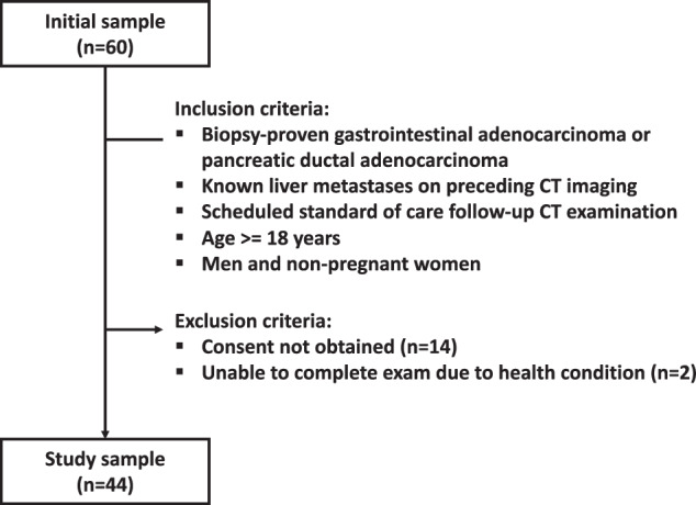
Materials and methods: Participants with known liver metastases from gastrointestinal and pancreatic adenocarcinoma were prospectively included from routine follow-up (October 2020 to March 2022). Participants received standard-dose CT and two additional reduced-dose scans during the same contrast administration, each reconstructed with IR and high-strength DLR. Two radiologists evaluated images for the presence of liver lesions, and a third established a reference standard. Diagnostic performance was compared using McNemar's test and mixed effects logistic regression.
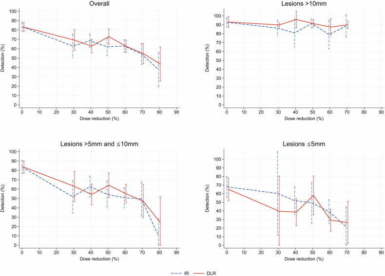
Results: Forty-four participants (mean age 66 years ± 11 [standard deviation], 28 men) were evaluated with 348 included liver lesions ≤ 20 mm (297 metastases, 51 benign; mean size 9.1 ± 4.3 mm). Mean volume CT dose index was 14.2, 7.8 mGy, and 5.1 mGy. Between algorithms, no significant difference in lesion detection was observed within dose levels. Detection of 233 lesions ≤ 10 mm was deteriorated with lower dose levels despite DLR denoising, with 185 detected at standard-dose IR (79.4%; 95% CI: 73.5-84.3) vs 128 at medium-dose DLR (54.9%; 95% CI: 48.3-61.4; p < 0.001) and 105 at low-dose DLR (45.1%; 95% CI: 38.6-51.7; p < 0.001).
Conclusion: Diagnostic performance for liver lesion detection was comparable between algorithms. When the detection of smaller lesions is important, DLR did not facilitate substantial dose reduction.
Key points: Question Methods to reduce CT radiation dose are desirable in clinical practice, and DLR has shown promising image denoising capabilities. Findings Liver lesion detection was comparable for DLR and IR across dose levels, but detection of smaller lesions deteriorated with lower dose levels. Clinical relevance Although potent in image noise reduction, the diagnostic performance of DLR is comparable to IR at standard-dose and reduced-dose CT. Care must be taken in pursuit of dose reduction when the detection and characterization of smaller liver lesions are of clinical importance.
期刊介绍:
European Radiology (ER) continuously updates scientific knowledge in radiology by publication of strong original articles and state-of-the-art reviews written by leading radiologists. A well balanced combination of review articles, original papers, short communications from European radiological congresses and information on society matters makes ER an indispensable source for current information in this field.
This is the Journal of the European Society of Radiology, and the official journal of a number of societies.
From 2004-2008 supplements to European Radiology were published under its companion, European Radiology Supplements, ISSN 1613-3749.

 求助内容:
求助内容: 应助结果提醒方式:


