Mert Ocak, Ferhat Geneci, Bilge İpek Torun, Mehmet Fatih Şentürk, Emine Şebnem Kurşun Çakmak
{"title":"锥形束计算机与微计算机体层摄影测量面部管裂的比较。","authors":"Mert Ocak, Ferhat Geneci, Bilge İpek Torun, Mehmet Fatih Şentürk, Emine Şebnem Kurşun Çakmak","doi":"10.1186/s13005-025-00485-x","DOIUrl":null,"url":null,"abstract":"<p><strong>Background: </strong>Selecting the correct imaging technique for critical anatomical structures is essential in descriptive studies and for supporting clinical applications. Facial canal dehiscence poses a significant risk for iatrogenic facial nerve injuries during middle ear surgeries. Accurate imaging is critical for surgical planning and minimizing complications. Detection of facial canal openings in the clinic is performed using imaging methods such as high-resolution computed tomography (HRCT). Studies have shown that the sensitivity of this method is approximately 66%. A high-resolution, 3D imaging method was used to measure the sensitivity of HRCT in the most accurate way.</p><p><strong>Aims/objectives: </strong>This study aimed to compare two radiological methods for measuring facial canal dehiscence. Specifically, we compared cone-beam computed tomography (CBCT) with high-resolution micro-computed tomography (micro-CT).</p><p><strong>Materials and methods: </strong>Thirty-six temporal bone specimens without external defects were used. The specimens were scanned using both CBCT and micro-CT. The presence of facial canal dehiscence in the tympanic segment of the facial nerve (FN) was evaluated. A paired sample t-test was used for statistical analysis, with significance set at p < 0.05.</p><p><strong>Results: </strong>Facial canal dehiscence was detected in 10 bones on micro-CT images, while 26 bones appeared intact. In contrast, CBCT images showed dehiscence in 25 bones, with 11 bones intact. Additionally, the mean dehiscence width was 3.469 mm (range: 1.577-8.921 mm) in micro-CT images, compared to 1.279 mm (range: 0.670-9.354 mm) in CBCT images. In the 10 bones where dehiscence was identified by both methods, the average width of the dehiscence measured 5.347 mm (range: 1.840-9.354 mm) in the CBCT images. The difference in measurements between CBCT and micro-CT was statistically significant (p < 0.05).</p><p><strong>Conclusions and significance: </strong>The low resolution of CBCT was insufficient for visualizing the thin bony tissue lining the facial canal. These findings suggest that the frequency of facial canal dehiscence measured in preoperative CBCT images may be overestimated compared to actual anatomical conditions. These findings provide critical insights for preoperative evaluation and surgical planning in middle ear procedures.</p>","PeriodicalId":12994,"journal":{"name":"Head & Face Medicine","volume":"21 1","pages":"33"},"PeriodicalIF":2.4000,"publicationDate":"2025-04-28","publicationTypes":"Journal Article","fieldsOfStudy":null,"isOpenAccess":false,"openAccessPdf":"https://www.ncbi.nlm.nih.gov/pmc/articles/PMC12036292/pdf/","citationCount":"0","resultStr":"{\"title\":\"Comparison of cone beam-computed and micro-computed tomography data for measuring facial canal dehiscence.\",\"authors\":\"Mert Ocak, Ferhat Geneci, Bilge İpek Torun, Mehmet Fatih Şentürk, Emine Şebnem Kurşun Çakmak\",\"doi\":\"10.1186/s13005-025-00485-x\",\"DOIUrl\":null,\"url\":null,\"abstract\":\"<p><strong>Background: </strong>Selecting the correct imaging technique for critical anatomical structures is essential in descriptive studies and for supporting clinical applications. Facial canal dehiscence poses a significant risk for iatrogenic facial nerve injuries during middle ear surgeries. Accurate imaging is critical for surgical planning and minimizing complications. Detection of facial canal openings in the clinic is performed using imaging methods such as high-resolution computed tomography (HRCT). Studies have shown that the sensitivity of this method is approximately 66%. A high-resolution, 3D imaging method was used to measure the sensitivity of HRCT in the most accurate way.</p><p><strong>Aims/objectives: </strong>This study aimed to compare two radiological methods for measuring facial canal dehiscence. Specifically, we compared cone-beam computed tomography (CBCT) with high-resolution micro-computed tomography (micro-CT).</p><p><strong>Materials and methods: </strong>Thirty-six temporal bone specimens without external defects were used. The specimens were scanned using both CBCT and micro-CT. The presence of facial canal dehiscence in the tympanic segment of the facial nerve (FN) was evaluated. A paired sample t-test was used for statistical analysis, with significance set at p < 0.05.</p><p><strong>Results: </strong>Facial canal dehiscence was detected in 10 bones on micro-CT images, while 26 bones appeared intact. In contrast, CBCT images showed dehiscence in 25 bones, with 11 bones intact. Additionally, the mean dehiscence width was 3.469 mm (range: 1.577-8.921 mm) in micro-CT images, compared to 1.279 mm (range: 0.670-9.354 mm) in CBCT images. In the 10 bones where dehiscence was identified by both methods, the average width of the dehiscence measured 5.347 mm (range: 1.840-9.354 mm) in the CBCT images. The difference in measurements between CBCT and micro-CT was statistically significant (p < 0.05).</p><p><strong>Conclusions and significance: </strong>The low resolution of CBCT was insufficient for visualizing the thin bony tissue lining the facial canal. These findings suggest that the frequency of facial canal dehiscence measured in preoperative CBCT images may be overestimated compared to actual anatomical conditions. These findings provide critical insights for preoperative evaluation and surgical planning in middle ear procedures.</p>\",\"PeriodicalId\":12994,\"journal\":{\"name\":\"Head & Face Medicine\",\"volume\":\"21 1\",\"pages\":\"33\"},\"PeriodicalIF\":2.4000,\"publicationDate\":\"2025-04-28\",\"publicationTypes\":\"Journal Article\",\"fieldsOfStudy\":null,\"isOpenAccess\":false,\"openAccessPdf\":\"https://www.ncbi.nlm.nih.gov/pmc/articles/PMC12036292/pdf/\",\"citationCount\":\"0\",\"resultStr\":null,\"platform\":\"Semanticscholar\",\"paperid\":null,\"PeriodicalName\":\"Head & Face Medicine\",\"FirstCategoryId\":\"3\",\"ListUrlMain\":\"https://doi.org/10.1186/s13005-025-00485-x\",\"RegionNum\":2,\"RegionCategory\":\"医学\",\"ArticlePicture\":[],\"TitleCN\":null,\"AbstractTextCN\":null,\"PMCID\":null,\"EPubDate\":\"\",\"PubModel\":\"\",\"JCR\":\"Q2\",\"JCRName\":\"DENTISTRY, ORAL SURGERY & MEDICINE\",\"Score\":null,\"Total\":0}","platform":"Semanticscholar","paperid":null,"PeriodicalName":"Head & Face Medicine","FirstCategoryId":"3","ListUrlMain":"https://doi.org/10.1186/s13005-025-00485-x","RegionNum":2,"RegionCategory":"医学","ArticlePicture":[],"TitleCN":null,"AbstractTextCN":null,"PMCID":null,"EPubDate":"","PubModel":"","JCR":"Q2","JCRName":"DENTISTRY, ORAL SURGERY & MEDICINE","Score":null,"Total":0}
Comparison of cone beam-computed and micro-computed tomography data for measuring facial canal dehiscence.
Background: Selecting the correct imaging technique for critical anatomical structures is essential in descriptive studies and for supporting clinical applications. Facial canal dehiscence poses a significant risk for iatrogenic facial nerve injuries during middle ear surgeries. Accurate imaging is critical for surgical planning and minimizing complications. Detection of facial canal openings in the clinic is performed using imaging methods such as high-resolution computed tomography (HRCT). Studies have shown that the sensitivity of this method is approximately 66%. A high-resolution, 3D imaging method was used to measure the sensitivity of HRCT in the most accurate way.
Aims/objectives: This study aimed to compare two radiological methods for measuring facial canal dehiscence. Specifically, we compared cone-beam computed tomography (CBCT) with high-resolution micro-computed tomography (micro-CT).
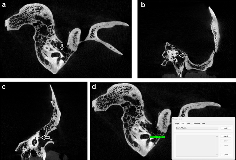
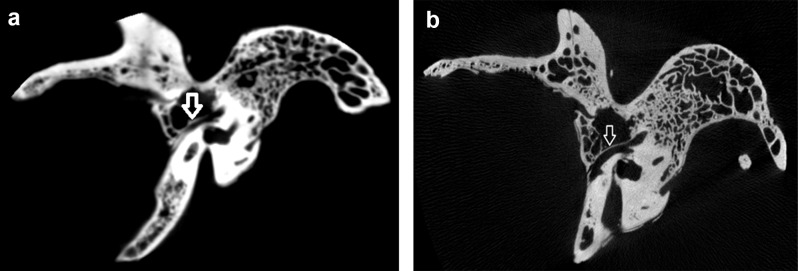
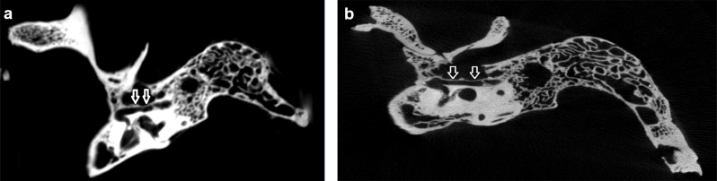
Materials and methods: Thirty-six temporal bone specimens without external defects were used. The specimens were scanned using both CBCT and micro-CT. The presence of facial canal dehiscence in the tympanic segment of the facial nerve (FN) was evaluated. A paired sample t-test was used for statistical analysis, with significance set at p < 0.05.
Results: Facial canal dehiscence was detected in 10 bones on micro-CT images, while 26 bones appeared intact. In contrast, CBCT images showed dehiscence in 25 bones, with 11 bones intact. Additionally, the mean dehiscence width was 3.469 mm (range: 1.577-8.921 mm) in micro-CT images, compared to 1.279 mm (range: 0.670-9.354 mm) in CBCT images. In the 10 bones where dehiscence was identified by both methods, the average width of the dehiscence measured 5.347 mm (range: 1.840-9.354 mm) in the CBCT images. The difference in measurements between CBCT and micro-CT was statistically significant (p < 0.05).
Conclusions and significance: The low resolution of CBCT was insufficient for visualizing the thin bony tissue lining the facial canal. These findings suggest that the frequency of facial canal dehiscence measured in preoperative CBCT images may be overestimated compared to actual anatomical conditions. These findings provide critical insights for preoperative evaluation and surgical planning in middle ear procedures.
期刊介绍:
Head & Face Medicine is a multidisciplinary open access journal that publishes basic and clinical research concerning all aspects of cranial, facial and oral conditions.
The journal covers all aspects of cranial, facial and oral diseases and their management. It has been designed as a multidisciplinary journal for clinicians and researchers involved in the diagnostic and therapeutic aspects of diseases which affect the human head and face. The journal is wide-ranging, covering the development, aetiology, epidemiology and therapy of head and face diseases to the basic science that underlies these diseases. Management of head and face diseases includes all aspects of surgical and non-surgical treatments including psychopharmacological therapies.
 求助内容:
求助内容: 应助结果提醒方式:


