{"title":"上颌窦评估:计算机断层分析和分类。","authors":"Mohammad Waheed El-Anwar, Mohamed Kamel Alawady, Hoda Ismail Abdelhamid, Tamer Oraby, Mohamed Talaat Albasiouny, Ashraf El-Hussiny","doi":"10.1055/s-0044-1791728","DOIUrl":null,"url":null,"abstract":"<p><p><b>Introduction</b> The preoperative assessment of the computed tomography (CT) characteristics of the maxillary sinus helps to preserve its anatomical and functional integrity during and after surgery. <b>Objective</b> To use CT scanning to identify maxillary sinus variations and types that were not previously published. <b>Methods</b> The present study was carried out on 110 paranasal CT scans (220 sides). Axial images were obtained with multiplanar scans, to visualize details in coronal and sagittal planes for all subjects. <b>Results</b> Among the 110 CTs (220 sides) of the maxillary sinus's floor, there were 53.2% type 1, 29.1% type 2, 10% type 3, and 7.7% type 4, with significant difference between genders. The most common maxillary sinus floor was type 1. The lateral maxillary sinus wall was found to be type 1 in 32.7%, type 2 in 65%, and type 3 in 2.3%, with a significant difference between genders. The most common lateral wall of the maxillary sinus type was type 2. The orbital floor was found to be type 1 in 0.9%, type 2 in 21.3%, type 3 in 50.5%, and type 4 in 27.3%, without significant difference between genders. Asymmetry was detected between the right and left sides for the maxillary sinus floor of in 22.7%, lateral maxillary wall in 16%, and orbital floor (maxillary roof) in 30%. <b>Conclusion</b> This study aims to increase surgeons' awareness of maxillary sinus variations, creating new classifications for usage and communication in the otorhinolaryngology and endoscopic fields. It could also be helpful for training medical residents.</p>","PeriodicalId":13731,"journal":{"name":"International Archives of Otorhinolaryngology","volume":"29 2","pages":"1-6"},"PeriodicalIF":1.1000,"publicationDate":"2025-04-15","publicationTypes":"Journal Article","fieldsOfStudy":null,"isOpenAccess":false,"openAccessPdf":"https://www.ncbi.nlm.nih.gov/pmc/articles/PMC12020587/pdf/","citationCount":"0","resultStr":"{\"title\":\"Maxillary Sinus Assessment: A Computed Tomography Analysis and Classification.\",\"authors\":\"Mohammad Waheed El-Anwar, Mohamed Kamel Alawady, Hoda Ismail Abdelhamid, Tamer Oraby, Mohamed Talaat Albasiouny, Ashraf El-Hussiny\",\"doi\":\"10.1055/s-0044-1791728\",\"DOIUrl\":null,\"url\":null,\"abstract\":\"<p><p><b>Introduction</b> The preoperative assessment of the computed tomography (CT) characteristics of the maxillary sinus helps to preserve its anatomical and functional integrity during and after surgery. <b>Objective</b> To use CT scanning to identify maxillary sinus variations and types that were not previously published. <b>Methods</b> The present study was carried out on 110 paranasal CT scans (220 sides). Axial images were obtained with multiplanar scans, to visualize details in coronal and sagittal planes for all subjects. <b>Results</b> Among the 110 CTs (220 sides) of the maxillary sinus's floor, there were 53.2% type 1, 29.1% type 2, 10% type 3, and 7.7% type 4, with significant difference between genders. The most common maxillary sinus floor was type 1. The lateral maxillary sinus wall was found to be type 1 in 32.7%, type 2 in 65%, and type 3 in 2.3%, with a significant difference between genders. The most common lateral wall of the maxillary sinus type was type 2. The orbital floor was found to be type 1 in 0.9%, type 2 in 21.3%, type 3 in 50.5%, and type 4 in 27.3%, without significant difference between genders. Asymmetry was detected between the right and left sides for the maxillary sinus floor of in 22.7%, lateral maxillary wall in 16%, and orbital floor (maxillary roof) in 30%. <b>Conclusion</b> This study aims to increase surgeons' awareness of maxillary sinus variations, creating new classifications for usage and communication in the otorhinolaryngology and endoscopic fields. It could also be helpful for training medical residents.</p>\",\"PeriodicalId\":13731,\"journal\":{\"name\":\"International Archives of Otorhinolaryngology\",\"volume\":\"29 2\",\"pages\":\"1-6\"},\"PeriodicalIF\":1.1000,\"publicationDate\":\"2025-04-15\",\"publicationTypes\":\"Journal Article\",\"fieldsOfStudy\":null,\"isOpenAccess\":false,\"openAccessPdf\":\"https://www.ncbi.nlm.nih.gov/pmc/articles/PMC12020587/pdf/\",\"citationCount\":\"0\",\"resultStr\":null,\"platform\":\"Semanticscholar\",\"paperid\":null,\"PeriodicalName\":\"International Archives of Otorhinolaryngology\",\"FirstCategoryId\":\"1085\",\"ListUrlMain\":\"https://doi.org/10.1055/s-0044-1791728\",\"RegionNum\":0,\"RegionCategory\":null,\"ArticlePicture\":[],\"TitleCN\":null,\"AbstractTextCN\":null,\"PMCID\":null,\"EPubDate\":\"2025/4/1 0:00:00\",\"PubModel\":\"eCollection\",\"JCR\":\"Q3\",\"JCRName\":\"OTORHINOLARYNGOLOGY\",\"Score\":null,\"Total\":0}","platform":"Semanticscholar","paperid":null,"PeriodicalName":"International Archives of Otorhinolaryngology","FirstCategoryId":"1085","ListUrlMain":"https://doi.org/10.1055/s-0044-1791728","RegionNum":0,"RegionCategory":null,"ArticlePicture":[],"TitleCN":null,"AbstractTextCN":null,"PMCID":null,"EPubDate":"2025/4/1 0:00:00","PubModel":"eCollection","JCR":"Q3","JCRName":"OTORHINOLARYNGOLOGY","Score":null,"Total":0}
Maxillary Sinus Assessment: A Computed Tomography Analysis and Classification.
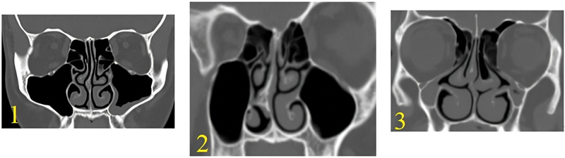
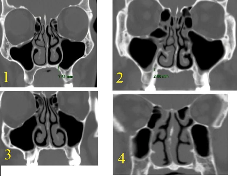
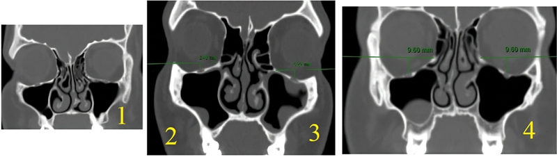
Introduction The preoperative assessment of the computed tomography (CT) characteristics of the maxillary sinus helps to preserve its anatomical and functional integrity during and after surgery. Objective To use CT scanning to identify maxillary sinus variations and types that were not previously published. Methods The present study was carried out on 110 paranasal CT scans (220 sides). Axial images were obtained with multiplanar scans, to visualize details in coronal and sagittal planes for all subjects. Results Among the 110 CTs (220 sides) of the maxillary sinus's floor, there were 53.2% type 1, 29.1% type 2, 10% type 3, and 7.7% type 4, with significant difference between genders. The most common maxillary sinus floor was type 1. The lateral maxillary sinus wall was found to be type 1 in 32.7%, type 2 in 65%, and type 3 in 2.3%, with a significant difference between genders. The most common lateral wall of the maxillary sinus type was type 2. The orbital floor was found to be type 1 in 0.9%, type 2 in 21.3%, type 3 in 50.5%, and type 4 in 27.3%, without significant difference between genders. Asymmetry was detected between the right and left sides for the maxillary sinus floor of in 22.7%, lateral maxillary wall in 16%, and orbital floor (maxillary roof) in 30%. Conclusion This study aims to increase surgeons' awareness of maxillary sinus variations, creating new classifications for usage and communication in the otorhinolaryngology and endoscopic fields. It could also be helpful for training medical residents.

 求助内容:
求助内容: 应助结果提醒方式:


