Xiaorui Wang, Sayo Maeno, Yixin Wang, Shizuka Koh, Shihao Chen, Andrew J Quantock, Siân R Morgan, Sally Hayes, Colm McAlinden
{"title":"角膜生物力学及OCT技术在圆锥角膜早期诊断中的应用。","authors":"Xiaorui Wang, Sayo Maeno, Yixin Wang, Shizuka Koh, Shihao Chen, Andrew J Quantock, Siân R Morgan, Sally Hayes, Colm McAlinden","doi":"10.1186/s40662-025-00435-3","DOIUrl":null,"url":null,"abstract":"<p><strong>Background: </strong>Early detection of keratoconus is essential for maximizing the potential of cross-linking treatments designed to halt keratoconus progression, minimizing the risks of iatrogenic ectasia as well as reducing the need for corneal transplantation. This review focuses on the progress that has been made in the early detection of keratoconus using biomechanical and topographical properties derived from three different technologies, namely the ocular response analyser (ORA), corneal visualization Scheimpflug tonometer (Corvis ST) and optical coherence tomography (OCT).</p><p><strong>Method: </strong>A PubMed search was performed using the keywords of 'early keratoconus', 'subclinical keratoconus', 'forme fruste keratoconus', 'very asymmetric ectasia with normal topography/tomography' and 'ocular response analyser' and/or 'Corvis ST'/'corneal visualized Scheimpflug tomographer/tomography' and/or 'optical coherence tomography/tomographer'.</p><p><strong>Results: </strong>The integration of biomechanical parameters and corneal morphological data from the topography/tomography or OCT, or the assessment of bilateral asymmetry, has demonstrated improvement in the accuracy of diagnosing early-stage keratoconus.</p><p><strong>Conclusions: </strong>As measurement principles differ depending on the technique used for keratoconus assessment, comprehensive metrics may be needed to reflect subtle anterior or posterior corneal changes and help identify eyes with very early ectasia. Although clinical experts have always, and will most likely, continue to play a pivotal role in decision-making for early keratoconus diagnosis, future developments in technology and AI may lead to enhanced early detection in the future.</p>","PeriodicalId":12194,"journal":{"name":"Eye and Vision","volume":"12 1","pages":"18"},"PeriodicalIF":4.0000,"publicationDate":"2025-05-12","publicationTypes":"Journal Article","fieldsOfStudy":null,"isOpenAccess":false,"openAccessPdf":"https://www.ncbi.nlm.nih.gov/pmc/articles/PMC12067920/pdf/","citationCount":"0","resultStr":"{\"title\":\"Early diagnosis of keratoconus using corneal biomechanics and OCT derived technologies.\",\"authors\":\"Xiaorui Wang, Sayo Maeno, Yixin Wang, Shizuka Koh, Shihao Chen, Andrew J Quantock, Siân R Morgan, Sally Hayes, Colm McAlinden\",\"doi\":\"10.1186/s40662-025-00435-3\",\"DOIUrl\":null,\"url\":null,\"abstract\":\"<p><strong>Background: </strong>Early detection of keratoconus is essential for maximizing the potential of cross-linking treatments designed to halt keratoconus progression, minimizing the risks of iatrogenic ectasia as well as reducing the need for corneal transplantation. This review focuses on the progress that has been made in the early detection of keratoconus using biomechanical and topographical properties derived from three different technologies, namely the ocular response analyser (ORA), corneal visualization Scheimpflug tonometer (Corvis ST) and optical coherence tomography (OCT).</p><p><strong>Method: </strong>A PubMed search was performed using the keywords of 'early keratoconus', 'subclinical keratoconus', 'forme fruste keratoconus', 'very asymmetric ectasia with normal topography/tomography' and 'ocular response analyser' and/or 'Corvis ST'/'corneal visualized Scheimpflug tomographer/tomography' and/or 'optical coherence tomography/tomographer'.</p><p><strong>Results: </strong>The integration of biomechanical parameters and corneal morphological data from the topography/tomography or OCT, or the assessment of bilateral asymmetry, has demonstrated improvement in the accuracy of diagnosing early-stage keratoconus.</p><p><strong>Conclusions: </strong>As measurement principles differ depending on the technique used for keratoconus assessment, comprehensive metrics may be needed to reflect subtle anterior or posterior corneal changes and help identify eyes with very early ectasia. Although clinical experts have always, and will most likely, continue to play a pivotal role in decision-making for early keratoconus diagnosis, future developments in technology and AI may lead to enhanced early detection in the future.</p>\",\"PeriodicalId\":12194,\"journal\":{\"name\":\"Eye and Vision\",\"volume\":\"12 1\",\"pages\":\"18\"},\"PeriodicalIF\":4.0000,\"publicationDate\":\"2025-05-12\",\"publicationTypes\":\"Journal Article\",\"fieldsOfStudy\":null,\"isOpenAccess\":false,\"openAccessPdf\":\"https://www.ncbi.nlm.nih.gov/pmc/articles/PMC12067920/pdf/\",\"citationCount\":\"0\",\"resultStr\":null,\"platform\":\"Semanticscholar\",\"paperid\":null,\"PeriodicalName\":\"Eye and Vision\",\"FirstCategoryId\":\"3\",\"ListUrlMain\":\"https://doi.org/10.1186/s40662-025-00435-3\",\"RegionNum\":1,\"RegionCategory\":\"医学\",\"ArticlePicture\":[],\"TitleCN\":null,\"AbstractTextCN\":null,\"PMCID\":null,\"EPubDate\":\"\",\"PubModel\":\"\",\"JCR\":\"Q1\",\"JCRName\":\"OPHTHALMOLOGY\",\"Score\":null,\"Total\":0}","platform":"Semanticscholar","paperid":null,"PeriodicalName":"Eye and Vision","FirstCategoryId":"3","ListUrlMain":"https://doi.org/10.1186/s40662-025-00435-3","RegionNum":1,"RegionCategory":"医学","ArticlePicture":[],"TitleCN":null,"AbstractTextCN":null,"PMCID":null,"EPubDate":"","PubModel":"","JCR":"Q1","JCRName":"OPHTHALMOLOGY","Score":null,"Total":0}
Early diagnosis of keratoconus using corneal biomechanics and OCT derived technologies.
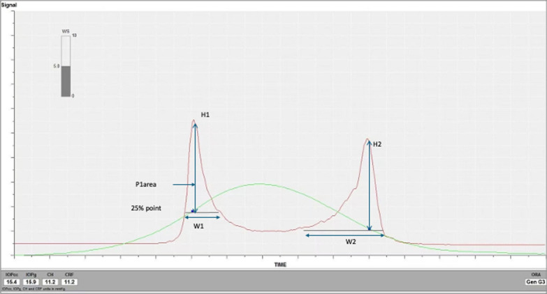
Background: Early detection of keratoconus is essential for maximizing the potential of cross-linking treatments designed to halt keratoconus progression, minimizing the risks of iatrogenic ectasia as well as reducing the need for corneal transplantation. This review focuses on the progress that has been made in the early detection of keratoconus using biomechanical and topographical properties derived from three different technologies, namely the ocular response analyser (ORA), corneal visualization Scheimpflug tonometer (Corvis ST) and optical coherence tomography (OCT).
Method: A PubMed search was performed using the keywords of 'early keratoconus', 'subclinical keratoconus', 'forme fruste keratoconus', 'very asymmetric ectasia with normal topography/tomography' and 'ocular response analyser' and/or 'Corvis ST'/'corneal visualized Scheimpflug tomographer/tomography' and/or 'optical coherence tomography/tomographer'.
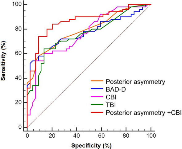
Results: The integration of biomechanical parameters and corneal morphological data from the topography/tomography or OCT, or the assessment of bilateral asymmetry, has demonstrated improvement in the accuracy of diagnosing early-stage keratoconus.
Conclusions: As measurement principles differ depending on the technique used for keratoconus assessment, comprehensive metrics may be needed to reflect subtle anterior or posterior corneal changes and help identify eyes with very early ectasia. Although clinical experts have always, and will most likely, continue to play a pivotal role in decision-making for early keratoconus diagnosis, future developments in technology and AI may lead to enhanced early detection in the future.
期刊介绍:
Eye and Vision is an open access, peer-reviewed journal for ophthalmologists and visual science specialists. It welcomes research articles, reviews, methodologies, commentaries, case reports, perspectives and short reports encompassing all aspects of eye and vision. Topics of interest include but are not limited to: current developments of theoretical, experimental and clinical investigations in ophthalmology, optometry and vision science which focus on novel and high-impact findings on central issues pertaining to biology, pathophysiology and etiology of eye diseases as well as advances in diagnostic techniques, surgical treatment, instrument updates, the latest drug findings, results of clinical trials and research findings. It aims to provide ophthalmologists and visual science specialists with the latest developments in theoretical, experimental and clinical investigations in eye and vision.
 求助内容:
求助内容: 应助结果提醒方式:


