Christian T Schamberger, Arnold J Suda, Tobias Grossner, Gerhard Schmidmaier, Stephan Stein
{"title":"基于超声的髋关节前头颈偏置的测定对于CAM畸形评估是可靠和可重复的。","authors":"Christian T Schamberger, Arnold J Suda, Tobias Grossner, Gerhard Schmidmaier, Stephan Stein","doi":"10.1055/a-2537-7181","DOIUrl":null,"url":null,"abstract":"<p><strong>Purpose: </strong>Native X-ray, magnetic resonance imaging (MRI), and computed tomography (CT) are standard methods for determining head-neck offset (HNO) in femoro-acetabular impingement (FAI). Our hypothesis was that sonography-assisted determination of the offset in CAM deformity of the hip is a cheap, radiation-free, and reliable alternative to conventional alpha-angle determination.</p><p><strong>Methods: </strong>Patients with hip pain and suspected CAM impingement who underwent anterior-longitudinal hip sonography according to DEGUM standard procedures and MRI were included in this single-center study between January 2015 and December 2019. Offset was determined three times on MRI and sonography by two independent investigators.</p><p><strong>Results: </strong>285 patients were screened and 110 patients (49 females, 61 males) met the inclusion criteria. The mean age at the time of investigation of 54 left and 56 right hip joints was 54.2 years. 1320 measurements were performed. No significant difference in HNO determination between MRI (6.11 mm+/-2.37) and sonography (5.93 mm+/-2.20) could be identified. The mean difference was 0.32 mm+/-0.32 mm (p>0.05) with a maximum deviation of 2.08 mm (outlier).</p><p><strong>Conclusion: </strong>Sonography-assisted determination of head-neck offset is a reliable and reproducible method and is not inferior to determination with MRI. Sonography can be used initially as an alternative or additional tool for the qualitative determination of CAM deformity of the hip joint.</p>","PeriodicalId":44852,"journal":{"name":"Ultrasound International Open","volume":"11 ","pages":"a25377181"},"PeriodicalIF":1.6000,"publicationDate":"2025-04-24","publicationTypes":"Journal Article","fieldsOfStudy":null,"isOpenAccess":false,"openAccessPdf":"https://www.ncbi.nlm.nih.gov/pmc/articles/PMC12039884/pdf/","citationCount":"0","resultStr":"{\"title\":\"Sonography-based determination of hip joint anterior head-neck offset is reliable and reproducible for CAM deformity assessment.\",\"authors\":\"Christian T Schamberger, Arnold J Suda, Tobias Grossner, Gerhard Schmidmaier, Stephan Stein\",\"doi\":\"10.1055/a-2537-7181\",\"DOIUrl\":null,\"url\":null,\"abstract\":\"<p><strong>Purpose: </strong>Native X-ray, magnetic resonance imaging (MRI), and computed tomography (CT) are standard methods for determining head-neck offset (HNO) in femoro-acetabular impingement (FAI). Our hypothesis was that sonography-assisted determination of the offset in CAM deformity of the hip is a cheap, radiation-free, and reliable alternative to conventional alpha-angle determination.</p><p><strong>Methods: </strong>Patients with hip pain and suspected CAM impingement who underwent anterior-longitudinal hip sonography according to DEGUM standard procedures and MRI were included in this single-center study between January 2015 and December 2019. Offset was determined three times on MRI and sonography by two independent investigators.</p><p><strong>Results: </strong>285 patients were screened and 110 patients (49 females, 61 males) met the inclusion criteria. The mean age at the time of investigation of 54 left and 56 right hip joints was 54.2 years. 1320 measurements were performed. No significant difference in HNO determination between MRI (6.11 mm+/-2.37) and sonography (5.93 mm+/-2.20) could be identified. The mean difference was 0.32 mm+/-0.32 mm (p>0.05) with a maximum deviation of 2.08 mm (outlier).</p><p><strong>Conclusion: </strong>Sonography-assisted determination of head-neck offset is a reliable and reproducible method and is not inferior to determination with MRI. Sonography can be used initially as an alternative or additional tool for the qualitative determination of CAM deformity of the hip joint.</p>\",\"PeriodicalId\":44852,\"journal\":{\"name\":\"Ultrasound International Open\",\"volume\":\"11 \",\"pages\":\"a25377181\"},\"PeriodicalIF\":1.6000,\"publicationDate\":\"2025-04-24\",\"publicationTypes\":\"Journal Article\",\"fieldsOfStudy\":null,\"isOpenAccess\":false,\"openAccessPdf\":\"https://www.ncbi.nlm.nih.gov/pmc/articles/PMC12039884/pdf/\",\"citationCount\":\"0\",\"resultStr\":null,\"platform\":\"Semanticscholar\",\"paperid\":null,\"PeriodicalName\":\"Ultrasound International Open\",\"FirstCategoryId\":\"1085\",\"ListUrlMain\":\"https://doi.org/10.1055/a-2537-7181\",\"RegionNum\":0,\"RegionCategory\":null,\"ArticlePicture\":[],\"TitleCN\":null,\"AbstractTextCN\":null,\"PMCID\":null,\"EPubDate\":\"2025/1/1 0:00:00\",\"PubModel\":\"eCollection\",\"JCR\":\"Q3\",\"JCRName\":\"RADIOLOGY, NUCLEAR MEDICINE & MEDICAL IMAGING\",\"Score\":null,\"Total\":0}","platform":"Semanticscholar","paperid":null,"PeriodicalName":"Ultrasound International Open","FirstCategoryId":"1085","ListUrlMain":"https://doi.org/10.1055/a-2537-7181","RegionNum":0,"RegionCategory":null,"ArticlePicture":[],"TitleCN":null,"AbstractTextCN":null,"PMCID":null,"EPubDate":"2025/1/1 0:00:00","PubModel":"eCollection","JCR":"Q3","JCRName":"RADIOLOGY, NUCLEAR MEDICINE & MEDICAL IMAGING","Score":null,"Total":0}
引用次数: 0
摘要
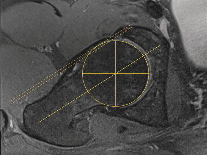
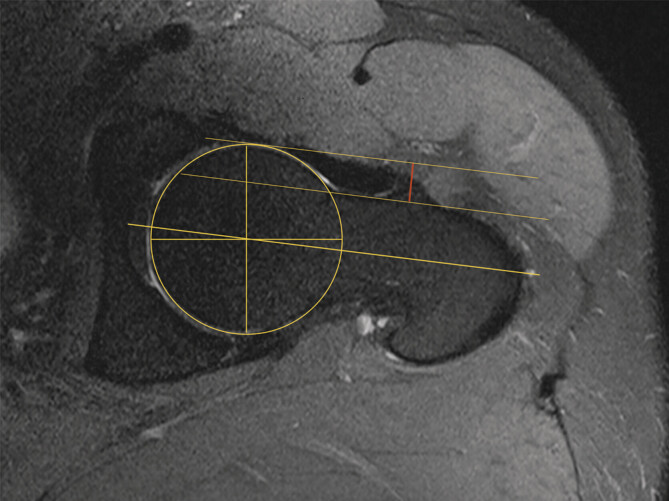
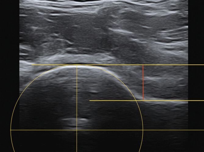
目的:x线,磁共振成像(MRI)和计算机断层扫描(CT)是确定股骨-髋臼撞击(FAI)头颈偏移(HNO)的标准方法。我们的假设是超声辅助确定髋关节CAM畸形的偏移量是一种便宜、无辐射、可靠的替代传统的α角测定方法。方法:在2015年1月至2019年12月期间,根据DEGUM标准程序和MRI进行前纵行髋关节超声检查的髋关节疼痛和疑似CAM撞击患者纳入本单中心研究。偏移量由两名独立调查员通过MRI和超声检查确定三次。结果:285例患者中,110例患者(女性49例,男性61例)符合纳入标准。54例左髋关节和56例右髋关节调查时的平均年龄为54.2岁。进行了1320次测量。MRI (6.11 mm+/-2.37)与超声(5.93 mm+/-2.20)检测HNO无显著差异。平均差值为0.32 mm±0.32 mm (p < 0.05),最大偏差为2.08 mm(异常值)。结论:超声辅助测定头颈偏移是一种可靠、可重复性高的方法,其准确性不低于MRI。超声检查最初可作为定性确定髋关节CAM畸形的替代或附加工具。
Sonography-based determination of hip joint anterior head-neck offset is reliable and reproducible for CAM deformity assessment.
Purpose: Native X-ray, magnetic resonance imaging (MRI), and computed tomography (CT) are standard methods for determining head-neck offset (HNO) in femoro-acetabular impingement (FAI). Our hypothesis was that sonography-assisted determination of the offset in CAM deformity of the hip is a cheap, radiation-free, and reliable alternative to conventional alpha-angle determination.
Methods: Patients with hip pain and suspected CAM impingement who underwent anterior-longitudinal hip sonography according to DEGUM standard procedures and MRI were included in this single-center study between January 2015 and December 2019. Offset was determined three times on MRI and sonography by two independent investigators.
Results: 285 patients were screened and 110 patients (49 females, 61 males) met the inclusion criteria. The mean age at the time of investigation of 54 left and 56 right hip joints was 54.2 years. 1320 measurements were performed. No significant difference in HNO determination between MRI (6.11 mm+/-2.37) and sonography (5.93 mm+/-2.20) could be identified. The mean difference was 0.32 mm+/-0.32 mm (p>0.05) with a maximum deviation of 2.08 mm (outlier).
Conclusion: Sonography-assisted determination of head-neck offset is a reliable and reproducible method and is not inferior to determination with MRI. Sonography can be used initially as an alternative or additional tool for the qualitative determination of CAM deformity of the hip joint.

 求助内容:
求助内容: 应助结果提醒方式:


