You-Sub Kim, Jae-Woong Kim, Sung-Pil Joo, Tae-Sun Kim
{"title":"抗胶粘剂替代及分步技术在减压颅骨切除术及后续颅骨成形术中的应用效果。","authors":"You-Sub Kim, Jae-Woong Kim, Sung-Pil Joo, Tae-Sun Kim","doi":"10.3340/jkns.2024.0176","DOIUrl":null,"url":null,"abstract":"<p><strong>Objective: </strong>Dural substitutes have been widely used in decompressive craniectomy to prevent adhesion, and have significantly reduced blood loss and operation time. However, there are only limited studies providing information regarding detailed techniques and the specific operation time that is associated with good prognoses. In this study, we evaluate the effectiveness of using a dural substitute as an anti-adhesive material during cranioplasty, focusing on technical details and operation time from incision to bone closure.</p><p><strong>Methods: </strong>A retrospectively reviewed total of 66 patients were included who underwent a craniectomy and subsequent cranioplasty caused by either a severe traumatic brain injury (n=35) or malignant infarction (n=31). The patients were divided into two groups depending on whether Neuro-Patch was used or not (31 in the Neuro-Patch group, 35 in the non-Neuro-Patch group). Propensity score matching was used to minimize the differences. Associated morbidities as well as operation time, and blood loss were analyzed and compared between the two groups.</p><p><strong>Results: </strong>To prevent adhesion, Neuro-Patch was placed as an onlay, enough to cover the surrounding skull at least 1 cm beyond the bone edges. A small piece was also placed over the temporalis muscle during the craniectomy. A step-by-step dissection was performed to minimize retraction-related injury during the subsequent cranioplasty. The mean estimated blood loss was significantly lower in the Neuro-Patch group (54.6±34.9 vs. 149.0±70.8 mL, p<0.001) and the mean time from incision to bone closure in the Neuro-Patch group was 40.8±14.3 minutes, which was significantly lower than in the non-Neuro-Patch group (91.5±38.2 minutes) as well. For each analysis of complications, the differences were not significant, however, the overall complication rate was significantly lower in the Neuro-Patch group (9.7%) than in the non-Neuro-Patch group (42.9%).</p><p><strong>Conclusion: </strong>Neuro-Patch can be used safely and effectively as an anti-adhesive substitute during cranioplasty. To improve clinical outcomes as well as intraoperative parameters including the time from incision to bone closure, planned placement of Neuro-Patch during craniectomy and the step-by-step dissection during cranioplasty is important.</p>","PeriodicalId":16283,"journal":{"name":"Journal of Korean Neurosurgical Society","volume":"68 3","pages":"360-368"},"PeriodicalIF":1.7000,"publicationDate":"2025-05-01","publicationTypes":"Journal Article","fieldsOfStudy":null,"isOpenAccess":false,"openAccessPdf":"https://www.ncbi.nlm.nih.gov/pmc/articles/PMC12062532/pdf/","citationCount":"0","resultStr":"{\"title\":\"Efficacy of Anti-Adhesive Substitute and Step-by-Step Techniques in Decompressive Craniectomy and Subsequent Cranioplasty.\",\"authors\":\"You-Sub Kim, Jae-Woong Kim, Sung-Pil Joo, Tae-Sun Kim\",\"doi\":\"10.3340/jkns.2024.0176\",\"DOIUrl\":null,\"url\":null,\"abstract\":\"<p><strong>Objective: </strong>Dural substitutes have been widely used in decompressive craniectomy to prevent adhesion, and have significantly reduced blood loss and operation time. However, there are only limited studies providing information regarding detailed techniques and the specific operation time that is associated with good prognoses. In this study, we evaluate the effectiveness of using a dural substitute as an anti-adhesive material during cranioplasty, focusing on technical details and operation time from incision to bone closure.</p><p><strong>Methods: </strong>A retrospectively reviewed total of 66 patients were included who underwent a craniectomy and subsequent cranioplasty caused by either a severe traumatic brain injury (n=35) or malignant infarction (n=31). The patients were divided into two groups depending on whether Neuro-Patch was used or not (31 in the Neuro-Patch group, 35 in the non-Neuro-Patch group). Propensity score matching was used to minimize the differences. Associated morbidities as well as operation time, and blood loss were analyzed and compared between the two groups.</p><p><strong>Results: </strong>To prevent adhesion, Neuro-Patch was placed as an onlay, enough to cover the surrounding skull at least 1 cm beyond the bone edges. A small piece was also placed over the temporalis muscle during the craniectomy. A step-by-step dissection was performed to minimize retraction-related injury during the subsequent cranioplasty. The mean estimated blood loss was significantly lower in the Neuro-Patch group (54.6±34.9 vs. 149.0±70.8 mL, p<0.001) and the mean time from incision to bone closure in the Neuro-Patch group was 40.8±14.3 minutes, which was significantly lower than in the non-Neuro-Patch group (91.5±38.2 minutes) as well. For each analysis of complications, the differences were not significant, however, the overall complication rate was significantly lower in the Neuro-Patch group (9.7%) than in the non-Neuro-Patch group (42.9%).</p><p><strong>Conclusion: </strong>Neuro-Patch can be used safely and effectively as an anti-adhesive substitute during cranioplasty. To improve clinical outcomes as well as intraoperative parameters including the time from incision to bone closure, planned placement of Neuro-Patch during craniectomy and the step-by-step dissection during cranioplasty is important.</p>\",\"PeriodicalId\":16283,\"journal\":{\"name\":\"Journal of Korean Neurosurgical Society\",\"volume\":\"68 3\",\"pages\":\"360-368\"},\"PeriodicalIF\":1.7000,\"publicationDate\":\"2025-05-01\",\"publicationTypes\":\"Journal Article\",\"fieldsOfStudy\":null,\"isOpenAccess\":false,\"openAccessPdf\":\"https://www.ncbi.nlm.nih.gov/pmc/articles/PMC12062532/pdf/\",\"citationCount\":\"0\",\"resultStr\":null,\"platform\":\"Semanticscholar\",\"paperid\":null,\"PeriodicalName\":\"Journal of Korean Neurosurgical Society\",\"FirstCategoryId\":\"3\",\"ListUrlMain\":\"https://doi.org/10.3340/jkns.2024.0176\",\"RegionNum\":4,\"RegionCategory\":\"医学\",\"ArticlePicture\":[],\"TitleCN\":null,\"AbstractTextCN\":null,\"PMCID\":null,\"EPubDate\":\"2025/4/11 0:00:00\",\"PubModel\":\"Epub\",\"JCR\":\"Q4\",\"JCRName\":\"CLINICAL NEUROLOGY\",\"Score\":null,\"Total\":0}","platform":"Semanticscholar","paperid":null,"PeriodicalName":"Journal of Korean Neurosurgical Society","FirstCategoryId":"3","ListUrlMain":"https://doi.org/10.3340/jkns.2024.0176","RegionNum":4,"RegionCategory":"医学","ArticlePicture":[],"TitleCN":null,"AbstractTextCN":null,"PMCID":null,"EPubDate":"2025/4/11 0:00:00","PubModel":"Epub","JCR":"Q4","JCRName":"CLINICAL NEUROLOGY","Score":null,"Total":0}
Efficacy of Anti-Adhesive Substitute and Step-by-Step Techniques in Decompressive Craniectomy and Subsequent Cranioplasty.
Objective: Dural substitutes have been widely used in decompressive craniectomy to prevent adhesion, and have significantly reduced blood loss and operation time. However, there are only limited studies providing information regarding detailed techniques and the specific operation time that is associated with good prognoses. In this study, we evaluate the effectiveness of using a dural substitute as an anti-adhesive material during cranioplasty, focusing on technical details and operation time from incision to bone closure.
Methods: A retrospectively reviewed total of 66 patients were included who underwent a craniectomy and subsequent cranioplasty caused by either a severe traumatic brain injury (n=35) or malignant infarction (n=31). The patients were divided into two groups depending on whether Neuro-Patch was used or not (31 in the Neuro-Patch group, 35 in the non-Neuro-Patch group). Propensity score matching was used to minimize the differences. Associated morbidities as well as operation time, and blood loss were analyzed and compared between the two groups.
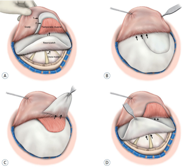
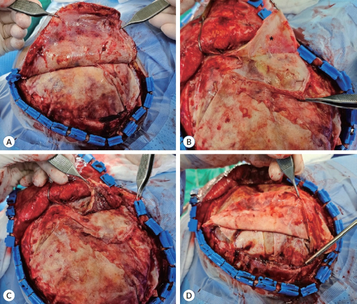
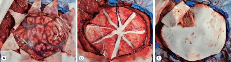
Results: To prevent adhesion, Neuro-Patch was placed as an onlay, enough to cover the surrounding skull at least 1 cm beyond the bone edges. A small piece was also placed over the temporalis muscle during the craniectomy. A step-by-step dissection was performed to minimize retraction-related injury during the subsequent cranioplasty. The mean estimated blood loss was significantly lower in the Neuro-Patch group (54.6±34.9 vs. 149.0±70.8 mL, p<0.001) and the mean time from incision to bone closure in the Neuro-Patch group was 40.8±14.3 minutes, which was significantly lower than in the non-Neuro-Patch group (91.5±38.2 minutes) as well. For each analysis of complications, the differences were not significant, however, the overall complication rate was significantly lower in the Neuro-Patch group (9.7%) than in the non-Neuro-Patch group (42.9%).
Conclusion: Neuro-Patch can be used safely and effectively as an anti-adhesive substitute during cranioplasty. To improve clinical outcomes as well as intraoperative parameters including the time from incision to bone closure, planned placement of Neuro-Patch during craniectomy and the step-by-step dissection during cranioplasty is important.
期刊介绍:
The Journal of Korean Neurosurgical Society (J Korean Neurosurg Soc) is the official journal of the Korean Neurosurgical Society, and published bimonthly (1st day of January, March, May, July, September, and November). It launched in October 31, 1972 with Volume 1 and Number 1. J Korean Neurosurg Soc aims to allow neurosurgeons from around the world to enrich their knowledge of patient management, education, and clinical or experimental research, and hence their professionalism. This journal publishes Laboratory Investigations, Clinical Articles, Review Articles, Case Reports, Technical Notes, and Letters to the Editor. Our field of interest involves clinical neurosurgery (cerebrovascular disease, neuro-oncology, skull base neurosurgery, spine, pediatric neurosurgery, functional neurosurgery, epilepsy, neuro-trauma, and peripheral nerve disease) and laboratory work in neuroscience.
 求助内容:
求助内容: 应助结果提醒方式:


