{"title":"探讨了EBT-XD薄膜剂量法标定时背景像素值的选择方法。","authors":"Sathiya Raj, Nithya Shree, Ganesh Kadirampatti","doi":"10.1002/pro6.1236","DOIUrl":null,"url":null,"abstract":"<p><strong>Purpose: </strong>This study investigates three different calibration methods for the selection of background pixel intensity.</p><p><strong>Methods: </strong>Film-by-Film (FBF) Method: Each film serves as its own control. Batch-by-Film (BBF) Method: A single film is used as a control for all calibration films. Generic (GEN) Method: A generic value (65535) is used as the background pixel value for all calibration films.Three calibration curves were established for the red, green, blue, and RGB channels, and the Radbard NIH (image) curve-fitting model was used to predict the dose. Sensitivity at different dose levels was quantified by calculating the first derivative of each color channel.</p><p><strong>Results: </strong>The GEN method exhibited a difference of up to 6% between the predicted and delivered doses below 2 Gy. The changes in optical density when using the GEN method differed significantly (<i>p</i><0.0001) from those of the FBF and BBF methods. In the dose range 5-30 Gy, the percentage difference between the predicted and delivered doses for the FBF, BBF, and GEN methods was within 2%. Both the red and green channels demonstrated higher sensitivity than the blue channel over the dose range of 2-30 Gy.</p><p><strong>Conclusions: </strong>The FBF method is more accurate than the BBF and GEN methods because it accounts for inter-film variations. The Radbard NIH (image) curve-fitting function proved suitable for predicting the dose for all the three calibration methods.</p>","PeriodicalId":32406,"journal":{"name":"Precision Radiation Oncology","volume":"8 3","pages":"132-137"},"PeriodicalIF":2.1000,"publicationDate":"2024-08-11","publicationTypes":"Journal Article","fieldsOfStudy":null,"isOpenAccess":false,"openAccessPdf":"https://www.ncbi.nlm.nih.gov/pmc/articles/PMC11935016/pdf/","citationCount":"0","resultStr":"{\"title\":\"Investigating the method of selection of background pixel values for the calibration of EBT-XD film dosimetry.\",\"authors\":\"Sathiya Raj, Nithya Shree, Ganesh Kadirampatti\",\"doi\":\"10.1002/pro6.1236\",\"DOIUrl\":null,\"url\":null,\"abstract\":\"<p><strong>Purpose: </strong>This study investigates three different calibration methods for the selection of background pixel intensity.</p><p><strong>Methods: </strong>Film-by-Film (FBF) Method: Each film serves as its own control. Batch-by-Film (BBF) Method: A single film is used as a control for all calibration films. Generic (GEN) Method: A generic value (65535) is used as the background pixel value for all calibration films.Three calibration curves were established for the red, green, blue, and RGB channels, and the Radbard NIH (image) curve-fitting model was used to predict the dose. Sensitivity at different dose levels was quantified by calculating the first derivative of each color channel.</p><p><strong>Results: </strong>The GEN method exhibited a difference of up to 6% between the predicted and delivered doses below 2 Gy. The changes in optical density when using the GEN method differed significantly (<i>p</i><0.0001) from those of the FBF and BBF methods. In the dose range 5-30 Gy, the percentage difference between the predicted and delivered doses for the FBF, BBF, and GEN methods was within 2%. Both the red and green channels demonstrated higher sensitivity than the blue channel over the dose range of 2-30 Gy.</p><p><strong>Conclusions: </strong>The FBF method is more accurate than the BBF and GEN methods because it accounts for inter-film variations. The Radbard NIH (image) curve-fitting function proved suitable for predicting the dose for all the three calibration methods.</p>\",\"PeriodicalId\":32406,\"journal\":{\"name\":\"Precision Radiation Oncology\",\"volume\":\"8 3\",\"pages\":\"132-137\"},\"PeriodicalIF\":2.1000,\"publicationDate\":\"2024-08-11\",\"publicationTypes\":\"Journal Article\",\"fieldsOfStudy\":null,\"isOpenAccess\":false,\"openAccessPdf\":\"https://www.ncbi.nlm.nih.gov/pmc/articles/PMC11935016/pdf/\",\"citationCount\":\"0\",\"resultStr\":null,\"platform\":\"Semanticscholar\",\"paperid\":null,\"PeriodicalName\":\"Precision Radiation Oncology\",\"FirstCategoryId\":\"1085\",\"ListUrlMain\":\"https://doi.org/10.1002/pro6.1236\",\"RegionNum\":0,\"RegionCategory\":null,\"ArticlePicture\":[],\"TitleCN\":null,\"AbstractTextCN\":null,\"PMCID\":null,\"EPubDate\":\"2024/9/1 0:00:00\",\"PubModel\":\"eCollection\",\"JCR\":\"Q4\",\"JCRName\":\"Medicine\",\"Score\":null,\"Total\":0}","platform":"Semanticscholar","paperid":null,"PeriodicalName":"Precision Radiation Oncology","FirstCategoryId":"1085","ListUrlMain":"https://doi.org/10.1002/pro6.1236","RegionNum":0,"RegionCategory":null,"ArticlePicture":[],"TitleCN":null,"AbstractTextCN":null,"PMCID":null,"EPubDate":"2024/9/1 0:00:00","PubModel":"eCollection","JCR":"Q4","JCRName":"Medicine","Score":null,"Total":0}
Investigating the method of selection of background pixel values for the calibration of EBT-XD film dosimetry.
Purpose: This study investigates three different calibration methods for the selection of background pixel intensity.
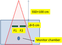
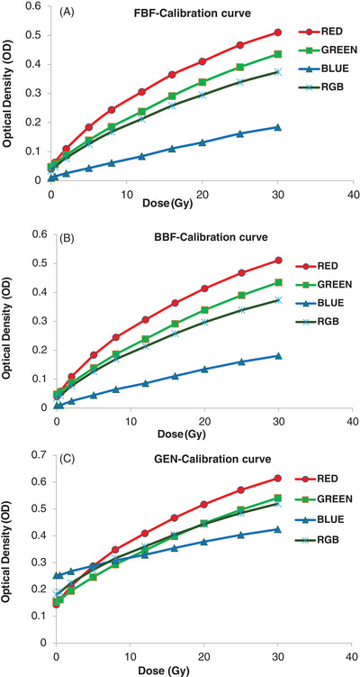
Methods: Film-by-Film (FBF) Method: Each film serves as its own control. Batch-by-Film (BBF) Method: A single film is used as a control for all calibration films. Generic (GEN) Method: A generic value (65535) is used as the background pixel value for all calibration films.Three calibration curves were established for the red, green, blue, and RGB channels, and the Radbard NIH (image) curve-fitting model was used to predict the dose. Sensitivity at different dose levels was quantified by calculating the first derivative of each color channel.
Results: The GEN method exhibited a difference of up to 6% between the predicted and delivered doses below 2 Gy. The changes in optical density when using the GEN method differed significantly (p<0.0001) from those of the FBF and BBF methods. In the dose range 5-30 Gy, the percentage difference between the predicted and delivered doses for the FBF, BBF, and GEN methods was within 2%. Both the red and green channels demonstrated higher sensitivity than the blue channel over the dose range of 2-30 Gy.
Conclusions: The FBF method is more accurate than the BBF and GEN methods because it accounts for inter-film variations. The Radbard NIH (image) curve-fitting function proved suitable for predicting the dose for all the three calibration methods.

 求助内容:
求助内容: 应助结果提醒方式:


