Cédric Dongmo Mayopa, David Ancelin, Pauline Despontin, Julie Manon, Gaspary Fodjeu, Randy Buzisa Mbuku, Maxime Masscheleyn, Olivier Cornu, Karim Tribak, Dan Putineanu
{"title":"Tillaux-Chaput结节骨折的预测因素:一项病例对照研究。","authors":"Cédric Dongmo Mayopa, David Ancelin, Pauline Despontin, Julie Manon, Gaspary Fodjeu, Randy Buzisa Mbuku, Maxime Masscheleyn, Olivier Cornu, Karim Tribak, Dan Putineanu","doi":"10.1177/24730114251332940","DOIUrl":null,"url":null,"abstract":"<p><strong>Background: </strong>Tillaux-Chaput tubercle fractures in adults often go unnoticed on standard radiographs in the setting of other malleolar fractures. This study aimed to identify clinical and radiologic factors associated with these fractures to develop a decision aid for computed tomographic (CT) diagnosis.</p><p><strong>Methods: </strong>This case-control study included 72 patients with bimalleolar fractures who underwent both radiography and CT scans. The case group consisted of 28 patients with Tillaux-Chaput fractures, and 44 served as control. Sociodemographic, clinical data, and injury mechanisms were compared using univariate and multivariate analysis to identify predictive factors.</p><p><strong>Results: </strong>Tillaux-Chaput fractures were undetected on standard radiographs in 60% of cases. In multivariate analysis, only age >60 years and injury mechanisms with Lauge-Hansen pronation-external rotation stage III or IV injuries were found to be highly associated with Tillaux-Chaput tubercle fractures.</p><p><strong>Conclusion: </strong>We recommend routine CT scan evaluation for patients >60 years old with stage III or IV pronation-external rotation ankle fractures.</p><p><strong>Level of evidence: </strong>Level III, case-control study.</p>","PeriodicalId":12429,"journal":{"name":"Foot & Ankle Orthopaedics","volume":"10 2","pages":"24730114251332940"},"PeriodicalIF":0.0000,"publicationDate":"2025-05-02","publicationTypes":"Journal Article","fieldsOfStudy":null,"isOpenAccess":false,"openAccessPdf":"https://www.ncbi.nlm.nih.gov/pmc/articles/PMC12049606/pdf/","citationCount":"0","resultStr":"{\"title\":\"Predictive Factors for Tillaux-Chaput Tubercle Fracture: A Case-Control Study.\",\"authors\":\"Cédric Dongmo Mayopa, David Ancelin, Pauline Despontin, Julie Manon, Gaspary Fodjeu, Randy Buzisa Mbuku, Maxime Masscheleyn, Olivier Cornu, Karim Tribak, Dan Putineanu\",\"doi\":\"10.1177/24730114251332940\",\"DOIUrl\":null,\"url\":null,\"abstract\":\"<p><strong>Background: </strong>Tillaux-Chaput tubercle fractures in adults often go unnoticed on standard radiographs in the setting of other malleolar fractures. This study aimed to identify clinical and radiologic factors associated with these fractures to develop a decision aid for computed tomographic (CT) diagnosis.</p><p><strong>Methods: </strong>This case-control study included 72 patients with bimalleolar fractures who underwent both radiography and CT scans. The case group consisted of 28 patients with Tillaux-Chaput fractures, and 44 served as control. Sociodemographic, clinical data, and injury mechanisms were compared using univariate and multivariate analysis to identify predictive factors.</p><p><strong>Results: </strong>Tillaux-Chaput fractures were undetected on standard radiographs in 60% of cases. In multivariate analysis, only age >60 years and injury mechanisms with Lauge-Hansen pronation-external rotation stage III or IV injuries were found to be highly associated with Tillaux-Chaput tubercle fractures.</p><p><strong>Conclusion: </strong>We recommend routine CT scan evaluation for patients >60 years old with stage III or IV pronation-external rotation ankle fractures.</p><p><strong>Level of evidence: </strong>Level III, case-control study.</p>\",\"PeriodicalId\":12429,\"journal\":{\"name\":\"Foot & Ankle Orthopaedics\",\"volume\":\"10 2\",\"pages\":\"24730114251332940\"},\"PeriodicalIF\":0.0000,\"publicationDate\":\"2025-05-02\",\"publicationTypes\":\"Journal Article\",\"fieldsOfStudy\":null,\"isOpenAccess\":false,\"openAccessPdf\":\"https://www.ncbi.nlm.nih.gov/pmc/articles/PMC12049606/pdf/\",\"citationCount\":\"0\",\"resultStr\":null,\"platform\":\"Semanticscholar\",\"paperid\":null,\"PeriodicalName\":\"Foot & Ankle Orthopaedics\",\"FirstCategoryId\":\"1085\",\"ListUrlMain\":\"https://doi.org/10.1177/24730114251332940\",\"RegionNum\":0,\"RegionCategory\":null,\"ArticlePicture\":[],\"TitleCN\":null,\"AbstractTextCN\":null,\"PMCID\":null,\"EPubDate\":\"2025/4/1 0:00:00\",\"PubModel\":\"eCollection\",\"JCR\":\"\",\"JCRName\":\"\",\"Score\":null,\"Total\":0}","platform":"Semanticscholar","paperid":null,"PeriodicalName":"Foot & Ankle Orthopaedics","FirstCategoryId":"1085","ListUrlMain":"https://doi.org/10.1177/24730114251332940","RegionNum":0,"RegionCategory":null,"ArticlePicture":[],"TitleCN":null,"AbstractTextCN":null,"PMCID":null,"EPubDate":"2025/4/1 0:00:00","PubModel":"eCollection","JCR":"","JCRName":"","Score":null,"Total":0}
Predictive Factors for Tillaux-Chaput Tubercle Fracture: A Case-Control Study.
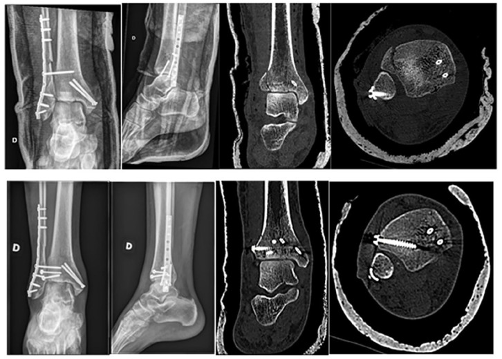
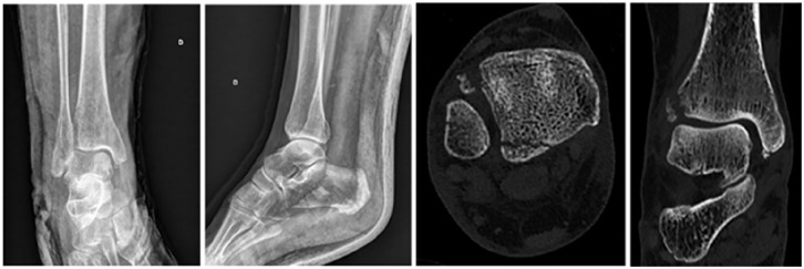
Background: Tillaux-Chaput tubercle fractures in adults often go unnoticed on standard radiographs in the setting of other malleolar fractures. This study aimed to identify clinical and radiologic factors associated with these fractures to develop a decision aid for computed tomographic (CT) diagnosis.
Methods: This case-control study included 72 patients with bimalleolar fractures who underwent both radiography and CT scans. The case group consisted of 28 patients with Tillaux-Chaput fractures, and 44 served as control. Sociodemographic, clinical data, and injury mechanisms were compared using univariate and multivariate analysis to identify predictive factors.
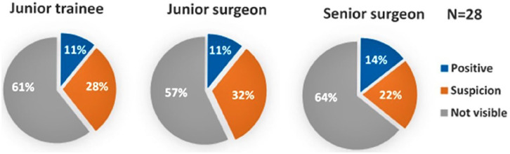
Results: Tillaux-Chaput fractures were undetected on standard radiographs in 60% of cases. In multivariate analysis, only age >60 years and injury mechanisms with Lauge-Hansen pronation-external rotation stage III or IV injuries were found to be highly associated with Tillaux-Chaput tubercle fractures.
Conclusion: We recommend routine CT scan evaluation for patients >60 years old with stage III or IV pronation-external rotation ankle fractures.

 求助内容:
求助内容: 应助结果提醒方式:


