Yassine N Azma, Nada Boci, Katarzyna Abramowicz, Luca Russo, Matthew R Orton, Nina Tunariu, Dow-Mu Koh, Geoffrey Charles-Edwards, David J Collins, Jessica M Winfield
{"title":"影像学方法对转移性前列腺癌骨髓脂肪含量评估的影响。","authors":"Yassine N Azma, Nada Boci, Katarzyna Abramowicz, Luca Russo, Matthew R Orton, Nina Tunariu, Dow-Mu Koh, Geoffrey Charles-Edwards, David J Collins, Jessica M Winfield","doi":"10.1007/s00330-025-11564-7","DOIUrl":null,"url":null,"abstract":"<p><strong>Objective: </strong>This study aimed to assess the accuracy of fat fraction estimation with clinically available Dixon sequences in normal-appearing marrow and bone metastases in the pelvis of metastatic prostate cancer patients.</p><p><strong>Methods: </strong>A prospective single-centre study was conducted with metastatic prostate cancer patients and healthy volunteers. Linearity and bias of fat fraction estimates from clinically available Dixon sequences were assessed against a 6-point PDw gradient echo (q-Dixon) sequence measuring the reference standard proton density fat fraction. Lesion fat fraction estimates were cross-compared using the Friedman test. Repeatability in volunteers was evaluated with Bland-Altman plots. Sensitivity of fat fraction estimates using TSE-Dixon sequences to specific absorption rate (SAR) related modifications were evaluated with correlation plots.</p><p><strong>Results: </strong>Thirty-three patients were recruited for this study. Significant (p < 0.05) absolute bias (12.4%) was demonstrated in the T1-weighted (T1w) Dixon measurements against the q-Dixon. Significant differences (p < 0.05) between fat fraction estimates provided by the T1w Dixon and PDw Dixon sequences were observed in 13 active and 6 treated lesions. Repeatability coefficients for fat fraction estimates ranged from 5.9 to 9.0% in the pelvic tissues of healthy volunteers. Reduction of slice number with repetition time for SAR had the greatest effect, reaching a maximum difference in fat fraction of 14.7% from the q-Dixon for the T2w-TSE Dixon in bone marrow.</p><p><strong>Conclusions: </strong>T1w Dixon methods can detect post-treatment changes but remain confounded by relaxation time biases. While all Dixon methods showed good repeatability, careful choice of SAR-related modifications is critical to maintaining accuracy for PD- and T2-weighted TSE sequences.</p><p><strong>Key points: </strong>Question The clinical validity of signal-weighted fat fraction estimates versus proton density fat fraction for characterising metastatic bone lesions has not been fully assessed. Findings T1-weighted Dixon sequences in line with whole-body MRI international guidelines demonstrate significant fat fraction bias, particularly in lesions and muscle. Clinical relevance Fat fraction estimation using T1-weighted Dixon sequences recommended in international guidelines are highly sensitive to relaxation time biases, making underlying physiological changes potentially ambiguous.</p>","PeriodicalId":12076,"journal":{"name":"European Radiology","volume":" ","pages":"6039-6051"},"PeriodicalIF":4.7000,"publicationDate":"2025-10-01","publicationTypes":"Journal Article","fieldsOfStudy":null,"isOpenAccess":false,"openAccessPdf":"https://www.ncbi.nlm.nih.gov/pmc/articles/PMC12417236/pdf/","citationCount":"0","resultStr":"{\"title\":\"Influence of imaging method on fat fraction estimation for assessing bone marrow in metastatic prostate cancer.\",\"authors\":\"Yassine N Azma, Nada Boci, Katarzyna Abramowicz, Luca Russo, Matthew R Orton, Nina Tunariu, Dow-Mu Koh, Geoffrey Charles-Edwards, David J Collins, Jessica M Winfield\",\"doi\":\"10.1007/s00330-025-11564-7\",\"DOIUrl\":null,\"url\":null,\"abstract\":\"<p><strong>Objective: </strong>This study aimed to assess the accuracy of fat fraction estimation with clinically available Dixon sequences in normal-appearing marrow and bone metastases in the pelvis of metastatic prostate cancer patients.</p><p><strong>Methods: </strong>A prospective single-centre study was conducted with metastatic prostate cancer patients and healthy volunteers. Linearity and bias of fat fraction estimates from clinically available Dixon sequences were assessed against a 6-point PDw gradient echo (q-Dixon) sequence measuring the reference standard proton density fat fraction. Lesion fat fraction estimates were cross-compared using the Friedman test. Repeatability in volunteers was evaluated with Bland-Altman plots. Sensitivity of fat fraction estimates using TSE-Dixon sequences to specific absorption rate (SAR) related modifications were evaluated with correlation plots.</p><p><strong>Results: </strong>Thirty-three patients were recruited for this study. Significant (p < 0.05) absolute bias (12.4%) was demonstrated in the T1-weighted (T1w) Dixon measurements against the q-Dixon. Significant differences (p < 0.05) between fat fraction estimates provided by the T1w Dixon and PDw Dixon sequences were observed in 13 active and 6 treated lesions. Repeatability coefficients for fat fraction estimates ranged from 5.9 to 9.0% in the pelvic tissues of healthy volunteers. Reduction of slice number with repetition time for SAR had the greatest effect, reaching a maximum difference in fat fraction of 14.7% from the q-Dixon for the T2w-TSE Dixon in bone marrow.</p><p><strong>Conclusions: </strong>T1w Dixon methods can detect post-treatment changes but remain confounded by relaxation time biases. While all Dixon methods showed good repeatability, careful choice of SAR-related modifications is critical to maintaining accuracy for PD- and T2-weighted TSE sequences.</p><p><strong>Key points: </strong>Question The clinical validity of signal-weighted fat fraction estimates versus proton density fat fraction for characterising metastatic bone lesions has not been fully assessed. Findings T1-weighted Dixon sequences in line with whole-body MRI international guidelines demonstrate significant fat fraction bias, particularly in lesions and muscle. Clinical relevance Fat fraction estimation using T1-weighted Dixon sequences recommended in international guidelines are highly sensitive to relaxation time biases, making underlying physiological changes potentially ambiguous.</p>\",\"PeriodicalId\":12076,\"journal\":{\"name\":\"European Radiology\",\"volume\":\" \",\"pages\":\"6039-6051\"},\"PeriodicalIF\":4.7000,\"publicationDate\":\"2025-10-01\",\"publicationTypes\":\"Journal Article\",\"fieldsOfStudy\":null,\"isOpenAccess\":false,\"openAccessPdf\":\"https://www.ncbi.nlm.nih.gov/pmc/articles/PMC12417236/pdf/\",\"citationCount\":\"0\",\"resultStr\":null,\"platform\":\"Semanticscholar\",\"paperid\":null,\"PeriodicalName\":\"European Radiology\",\"FirstCategoryId\":\"3\",\"ListUrlMain\":\"https://doi.org/10.1007/s00330-025-11564-7\",\"RegionNum\":2,\"RegionCategory\":\"医学\",\"ArticlePicture\":[],\"TitleCN\":null,\"AbstractTextCN\":null,\"PMCID\":null,\"EPubDate\":\"2025/4/11 0:00:00\",\"PubModel\":\"Epub\",\"JCR\":\"Q1\",\"JCRName\":\"RADIOLOGY, NUCLEAR MEDICINE & MEDICAL IMAGING\",\"Score\":null,\"Total\":0}","platform":"Semanticscholar","paperid":null,"PeriodicalName":"European Radiology","FirstCategoryId":"3","ListUrlMain":"https://doi.org/10.1007/s00330-025-11564-7","RegionNum":2,"RegionCategory":"医学","ArticlePicture":[],"TitleCN":null,"AbstractTextCN":null,"PMCID":null,"EPubDate":"2025/4/11 0:00:00","PubModel":"Epub","JCR":"Q1","JCRName":"RADIOLOGY, NUCLEAR MEDICINE & MEDICAL IMAGING","Score":null,"Total":0}
Influence of imaging method on fat fraction estimation for assessing bone marrow in metastatic prostate cancer.
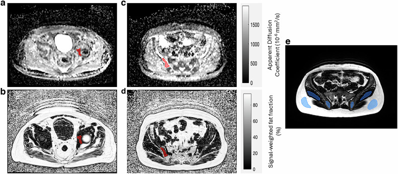
Objective: This study aimed to assess the accuracy of fat fraction estimation with clinically available Dixon sequences in normal-appearing marrow and bone metastases in the pelvis of metastatic prostate cancer patients.
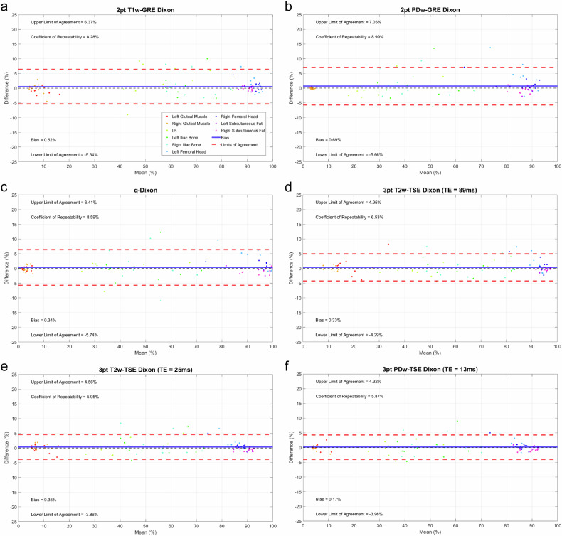
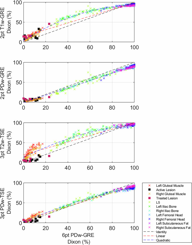
Methods: A prospective single-centre study was conducted with metastatic prostate cancer patients and healthy volunteers. Linearity and bias of fat fraction estimates from clinically available Dixon sequences were assessed against a 6-point PDw gradient echo (q-Dixon) sequence measuring the reference standard proton density fat fraction. Lesion fat fraction estimates were cross-compared using the Friedman test. Repeatability in volunteers was evaluated with Bland-Altman plots. Sensitivity of fat fraction estimates using TSE-Dixon sequences to specific absorption rate (SAR) related modifications were evaluated with correlation plots.
Results: Thirty-three patients were recruited for this study. Significant (p < 0.05) absolute bias (12.4%) was demonstrated in the T1-weighted (T1w) Dixon measurements against the q-Dixon. Significant differences (p < 0.05) between fat fraction estimates provided by the T1w Dixon and PDw Dixon sequences were observed in 13 active and 6 treated lesions. Repeatability coefficients for fat fraction estimates ranged from 5.9 to 9.0% in the pelvic tissues of healthy volunteers. Reduction of slice number with repetition time for SAR had the greatest effect, reaching a maximum difference in fat fraction of 14.7% from the q-Dixon for the T2w-TSE Dixon in bone marrow.
Conclusions: T1w Dixon methods can detect post-treatment changes but remain confounded by relaxation time biases. While all Dixon methods showed good repeatability, careful choice of SAR-related modifications is critical to maintaining accuracy for PD- and T2-weighted TSE sequences.
Key points: Question The clinical validity of signal-weighted fat fraction estimates versus proton density fat fraction for characterising metastatic bone lesions has not been fully assessed. Findings T1-weighted Dixon sequences in line with whole-body MRI international guidelines demonstrate significant fat fraction bias, particularly in lesions and muscle. Clinical relevance Fat fraction estimation using T1-weighted Dixon sequences recommended in international guidelines are highly sensitive to relaxation time biases, making underlying physiological changes potentially ambiguous.
期刊介绍:
European Radiology (ER) continuously updates scientific knowledge in radiology by publication of strong original articles and state-of-the-art reviews written by leading radiologists. A well balanced combination of review articles, original papers, short communications from European radiological congresses and information on society matters makes ER an indispensable source for current information in this field.
This is the Journal of the European Society of Radiology, and the official journal of a number of societies.
From 2004-2008 supplements to European Radiology were published under its companion, European Radiology Supplements, ISSN 1613-3749.

 求助内容:
求助内容: 应助结果提醒方式:


