{"title":"家族性腺瘤性息肉病偶遇圆顶型腺癌1例报告及文献复习。","authors":"Ying-Ying Chang, Xiao-Long Zhang, Yao-Hui Wang, Ting-Sheng Ling","doi":"10.1186/s13000-025-01633-2","DOIUrl":null,"url":null,"abstract":"<p><p>Dome-type carcinoma (DC), also referred as Gut-associated lymphoid tissue (GALT) carcinoma, is a rare variant of colorectal adenocarcinoma which has been seldomly reported up to now. We report a case of a DC lesion developed in a 33-year-old male diagnosed with family adenomatous polyposis (FAP). A 1.5 × 1.5 cm well-demarcated lesion exhibited a 0-Is + IIc figure was detected near the anastomotic stoma during regular colonoscopic polypectomy. Surgical specimen showed well-differentiated adenocarcinoma consisted of dilated cystic glands and the lymphoid stroma with reactive germinal centers exhibited a destructive manner of infiltration into SM2 level. The immunohistochemical findings revealed MUC1 positive but MUC2 negative of the carcinomas epithelial which retained all the 4 mismatch repair proteins (MMRs) (MLH1, PMS2, MSH2, and MSH6) and was negative for EBV-encoded small RNA-1 (EBER). Considered a rare category of colorectal adenocarcinoma, more cases will help uncover the nature of GALT/dome-type carcinoma. Clinicians and pathologists should be aware of recognizing this special type of carcinoma and making necessary differential diagnostics.</p>","PeriodicalId":11237,"journal":{"name":"Diagnostic Pathology","volume":"20 1","pages":"47"},"PeriodicalIF":2.3000,"publicationDate":"2025-04-17","publicationTypes":"Journal Article","fieldsOfStudy":null,"isOpenAccess":false,"openAccessPdf":"https://www.ncbi.nlm.nih.gov/pmc/articles/PMC12004612/pdf/","citationCount":"0","resultStr":"{\"title\":\"Family adenomatous polyposis come across dome type adenocarcinoma: a case report and literature review.\",\"authors\":\"Ying-Ying Chang, Xiao-Long Zhang, Yao-Hui Wang, Ting-Sheng Ling\",\"doi\":\"10.1186/s13000-025-01633-2\",\"DOIUrl\":null,\"url\":null,\"abstract\":\"<p><p>Dome-type carcinoma (DC), also referred as Gut-associated lymphoid tissue (GALT) carcinoma, is a rare variant of colorectal adenocarcinoma which has been seldomly reported up to now. We report a case of a DC lesion developed in a 33-year-old male diagnosed with family adenomatous polyposis (FAP). A 1.5 × 1.5 cm well-demarcated lesion exhibited a 0-Is + IIc figure was detected near the anastomotic stoma during regular colonoscopic polypectomy. Surgical specimen showed well-differentiated adenocarcinoma consisted of dilated cystic glands and the lymphoid stroma with reactive germinal centers exhibited a destructive manner of infiltration into SM2 level. The immunohistochemical findings revealed MUC1 positive but MUC2 negative of the carcinomas epithelial which retained all the 4 mismatch repair proteins (MMRs) (MLH1, PMS2, MSH2, and MSH6) and was negative for EBV-encoded small RNA-1 (EBER). Considered a rare category of colorectal adenocarcinoma, more cases will help uncover the nature of GALT/dome-type carcinoma. Clinicians and pathologists should be aware of recognizing this special type of carcinoma and making necessary differential diagnostics.</p>\",\"PeriodicalId\":11237,\"journal\":{\"name\":\"Diagnostic Pathology\",\"volume\":\"20 1\",\"pages\":\"47\"},\"PeriodicalIF\":2.3000,\"publicationDate\":\"2025-04-17\",\"publicationTypes\":\"Journal Article\",\"fieldsOfStudy\":null,\"isOpenAccess\":false,\"openAccessPdf\":\"https://www.ncbi.nlm.nih.gov/pmc/articles/PMC12004612/pdf/\",\"citationCount\":\"0\",\"resultStr\":null,\"platform\":\"Semanticscholar\",\"paperid\":null,\"PeriodicalName\":\"Diagnostic Pathology\",\"FirstCategoryId\":\"3\",\"ListUrlMain\":\"https://doi.org/10.1186/s13000-025-01633-2\",\"RegionNum\":3,\"RegionCategory\":\"医学\",\"ArticlePicture\":[],\"TitleCN\":null,\"AbstractTextCN\":null,\"PMCID\":null,\"EPubDate\":\"\",\"PubModel\":\"\",\"JCR\":\"Q2\",\"JCRName\":\"PATHOLOGY\",\"Score\":null,\"Total\":0}","platform":"Semanticscholar","paperid":null,"PeriodicalName":"Diagnostic Pathology","FirstCategoryId":"3","ListUrlMain":"https://doi.org/10.1186/s13000-025-01633-2","RegionNum":3,"RegionCategory":"医学","ArticlePicture":[],"TitleCN":null,"AbstractTextCN":null,"PMCID":null,"EPubDate":"","PubModel":"","JCR":"Q2","JCRName":"PATHOLOGY","Score":null,"Total":0}
Family adenomatous polyposis come across dome type adenocarcinoma: a case report and literature review.
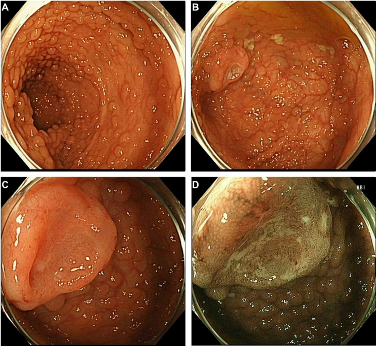
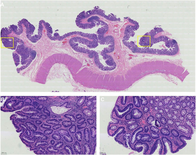
Dome-type carcinoma (DC), also referred as Gut-associated lymphoid tissue (GALT) carcinoma, is a rare variant of colorectal adenocarcinoma which has been seldomly reported up to now. We report a case of a DC lesion developed in a 33-year-old male diagnosed with family adenomatous polyposis (FAP). A 1.5 × 1.5 cm well-demarcated lesion exhibited a 0-Is + IIc figure was detected near the anastomotic stoma during regular colonoscopic polypectomy. Surgical specimen showed well-differentiated adenocarcinoma consisted of dilated cystic glands and the lymphoid stroma with reactive germinal centers exhibited a destructive manner of infiltration into SM2 level. The immunohistochemical findings revealed MUC1 positive but MUC2 negative of the carcinomas epithelial which retained all the 4 mismatch repair proteins (MMRs) (MLH1, PMS2, MSH2, and MSH6) and was negative for EBV-encoded small RNA-1 (EBER). Considered a rare category of colorectal adenocarcinoma, more cases will help uncover the nature of GALT/dome-type carcinoma. Clinicians and pathologists should be aware of recognizing this special type of carcinoma and making necessary differential diagnostics.
期刊介绍:
Diagnostic Pathology is an open access, peer-reviewed, online journal that considers research in surgical and clinical pathology, immunology, and biology, with a special focus on cutting-edge approaches in diagnostic pathology and tissue-based therapy. The journal covers all aspects of surgical pathology, including classic diagnostic pathology, prognosis-related diagnosis (tumor stages, prognosis markers, such as MIB-percentage, hormone receptors, etc.), and therapy-related findings. The journal also focuses on the technological aspects of pathology, including molecular biology techniques, morphometry aspects (stereology, DNA analysis, syntactic structure analysis), communication aspects (telecommunication, virtual microscopy, virtual pathology institutions, etc.), and electronic education and quality assurance (for example interactive publication, on-line references with automated updating, etc.).

 求助内容:
求助内容: 应助结果提醒方式:


