Haris Omić, Farsad Eskandary, Dietrich Beitzke, Marcos Wolf, Nicolas Kozakowski, Georg Böhmig, Andrea Beck-Tölly, Michael Eder
{"title":"T1松弛时间对肾移植功能障碍的预测。","authors":"Haris Omić, Farsad Eskandary, Dietrich Beitzke, Marcos Wolf, Nicolas Kozakowski, Georg Böhmig, Andrea Beck-Tölly, Michael Eder","doi":"10.3389/ti.2025.14301","DOIUrl":null,"url":null,"abstract":"<p><p>Quantitative magnetic resonance imaging (MRI) is emerging as a non-invasive tool to measure tissue scarring in renal allografts. However, whether prolonged T<sub>1</sub> relaxation time results in lower transplant survival rates is unknown. This retrospective cohort study analyzed the capability to predict renal allograft dysfunction based on median T<sub>1</sub> time. Forty-six transplant recipients with non-contrast 1.5T MRI and allograft biopsy were included. The primary endpoint was the eGFR slope over 24 months. T<sub>1</sub> relaxation time correlated significantly with eGFR levels at all follow-up stages. Patients with T<sub>1</sub> relaxation time above the median (T<sub>1</sub> <sup>high</sup>) had a consistent decline in kidney function as compared to the patient group below the median (T<sub>1</sub> <sup>low</sup>): overall eGFR slope: 11.3 vs. 1.4 mL/min/1.73 m<sup>2</sup> over 24 months, p = 0.016. Graft survival rates at 24 months were 52% in the T<sub>1</sub> <sup>high</sup> vs. 87% in the T<sub>1</sub> <sup>low</sup> group, p = 0.0015. ROC analysis discovered a positive predictive value of 52% and a negative predictive value of 91% for graft loss. T<sub>1</sub> mapping identified patients with a persistent decline of allograft function and an increased risk of allograft loss. MRI could significantly influence monitoring strategies in transplant surveillance, offering a safe, non-invasive alternative to traditional diagnostic methods.</p>","PeriodicalId":23343,"journal":{"name":"Transplant International","volume":"38 ","pages":"14301"},"PeriodicalIF":3.0000,"publicationDate":"2025-04-10","publicationTypes":"Journal Article","fieldsOfStudy":null,"isOpenAccess":false,"openAccessPdf":"https://www.ncbi.nlm.nih.gov/pmc/articles/PMC12018245/pdf/","citationCount":"0","resultStr":"{\"title\":\"T<sub>1</sub> Relaxation Time for the Prediction of Renal Transplant Dysfunction.\",\"authors\":\"Haris Omić, Farsad Eskandary, Dietrich Beitzke, Marcos Wolf, Nicolas Kozakowski, Georg Böhmig, Andrea Beck-Tölly, Michael Eder\",\"doi\":\"10.3389/ti.2025.14301\",\"DOIUrl\":null,\"url\":null,\"abstract\":\"<p><p>Quantitative magnetic resonance imaging (MRI) is emerging as a non-invasive tool to measure tissue scarring in renal allografts. However, whether prolonged T<sub>1</sub> relaxation time results in lower transplant survival rates is unknown. This retrospective cohort study analyzed the capability to predict renal allograft dysfunction based on median T<sub>1</sub> time. Forty-six transplant recipients with non-contrast 1.5T MRI and allograft biopsy were included. The primary endpoint was the eGFR slope over 24 months. T<sub>1</sub> relaxation time correlated significantly with eGFR levels at all follow-up stages. Patients with T<sub>1</sub> relaxation time above the median (T<sub>1</sub> <sup>high</sup>) had a consistent decline in kidney function as compared to the patient group below the median (T<sub>1</sub> <sup>low</sup>): overall eGFR slope: 11.3 vs. 1.4 mL/min/1.73 m<sup>2</sup> over 24 months, p = 0.016. Graft survival rates at 24 months were 52% in the T<sub>1</sub> <sup>high</sup> vs. 87% in the T<sub>1</sub> <sup>low</sup> group, p = 0.0015. ROC analysis discovered a positive predictive value of 52% and a negative predictive value of 91% for graft loss. T<sub>1</sub> mapping identified patients with a persistent decline of allograft function and an increased risk of allograft loss. MRI could significantly influence monitoring strategies in transplant surveillance, offering a safe, non-invasive alternative to traditional diagnostic methods.</p>\",\"PeriodicalId\":23343,\"journal\":{\"name\":\"Transplant International\",\"volume\":\"38 \",\"pages\":\"14301\"},\"PeriodicalIF\":3.0000,\"publicationDate\":\"2025-04-10\",\"publicationTypes\":\"Journal Article\",\"fieldsOfStudy\":null,\"isOpenAccess\":false,\"openAccessPdf\":\"https://www.ncbi.nlm.nih.gov/pmc/articles/PMC12018245/pdf/\",\"citationCount\":\"0\",\"resultStr\":null,\"platform\":\"Semanticscholar\",\"paperid\":null,\"PeriodicalName\":\"Transplant International\",\"FirstCategoryId\":\"3\",\"ListUrlMain\":\"https://doi.org/10.3389/ti.2025.14301\",\"RegionNum\":3,\"RegionCategory\":\"医学\",\"ArticlePicture\":[],\"TitleCN\":null,\"AbstractTextCN\":null,\"PMCID\":null,\"EPubDate\":\"2025/1/1 0:00:00\",\"PubModel\":\"eCollection\",\"JCR\":\"Q1\",\"JCRName\":\"SURGERY\",\"Score\":null,\"Total\":0}","platform":"Semanticscholar","paperid":null,"PeriodicalName":"Transplant International","FirstCategoryId":"3","ListUrlMain":"https://doi.org/10.3389/ti.2025.14301","RegionNum":3,"RegionCategory":"医学","ArticlePicture":[],"TitleCN":null,"AbstractTextCN":null,"PMCID":null,"EPubDate":"2025/1/1 0:00:00","PubModel":"eCollection","JCR":"Q1","JCRName":"SURGERY","Score":null,"Total":0}
引用次数: 0
摘要
定量磁共振成像(MRI)正在成为一种测量同种异体肾移植组织瘢痕形成的非侵入性工具。然而,延长T1松弛时间是否会导致移植存活率降低尚不清楚。本回顾性队列研究分析了基于中位T1时间预测同种异体肾移植功能障碍的能力。46例移植受者行非对比1.5T MRI和同种异体移植活检。主要终点是超过24个月的eGFR斜率。T1松弛时间与随访各阶段eGFR水平显著相关。与T1松弛时间低于中位数(T1低)的患者组相比,T1松弛时间高于中位数(T1高)的患者肾功能持续下降:在24个月内,总体eGFR斜率:11.3 vs. 1.4 mL/min/1.73 m2, p = 0.016。T1高组24个月移植物存活率为52%,T1低组为87%,p = 0.0015。ROC分析发现移植物损失的阳性预测值为52%,阴性预测值为91%。T1图谱确定了同种异体移植物功能持续下降和同种异体移植物丧失风险增加的患者。MRI可以显著影响移植监测的监测策略,为传统诊断方法提供了一种安全、无创的替代方法。
T1 Relaxation Time for the Prediction of Renal Transplant Dysfunction.
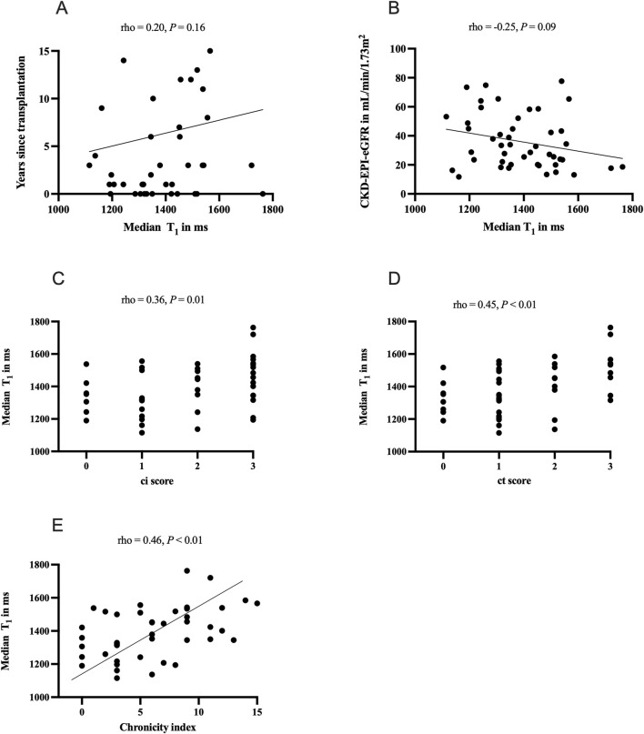
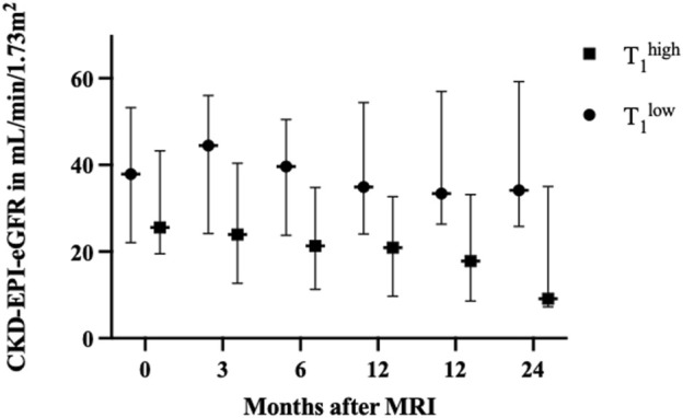
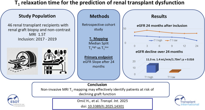
Quantitative magnetic resonance imaging (MRI) is emerging as a non-invasive tool to measure tissue scarring in renal allografts. However, whether prolonged T1 relaxation time results in lower transplant survival rates is unknown. This retrospective cohort study analyzed the capability to predict renal allograft dysfunction based on median T1 time. Forty-six transplant recipients with non-contrast 1.5T MRI and allograft biopsy were included. The primary endpoint was the eGFR slope over 24 months. T1 relaxation time correlated significantly with eGFR levels at all follow-up stages. Patients with T1 relaxation time above the median (T1high) had a consistent decline in kidney function as compared to the patient group below the median (T1low): overall eGFR slope: 11.3 vs. 1.4 mL/min/1.73 m2 over 24 months, p = 0.016. Graft survival rates at 24 months were 52% in the T1high vs. 87% in the T1low group, p = 0.0015. ROC analysis discovered a positive predictive value of 52% and a negative predictive value of 91% for graft loss. T1 mapping identified patients with a persistent decline of allograft function and an increased risk of allograft loss. MRI could significantly influence monitoring strategies in transplant surveillance, offering a safe, non-invasive alternative to traditional diagnostic methods.
期刊介绍:
The aim of the journal is to serve as a forum for the exchange of scientific information in the form of original and high quality papers in the field of transplantation. Clinical and experimental studies, as well as editorials, letters to the editors, and, occasionally, reviews on the biology, physiology, and immunology of transplantation of tissues and organs, are published. Publishing time for the latter is approximately six months, provided major revisions are not needed. The journal is published in yearly volumes, each volume containing twelve issues. Papers submitted to the journal are subject to peer review.
 求助内容:
求助内容: 应助结果提醒方式:


