Anson W Wilks, Kiana M Vakil-Gilani, William D Rooney, Dongseok Choi, Daniela Ghetie, Nizar Chahin
{"title":"免疫介导的坏死性肌病和皮肌炎中大腿肌肉受累的MRI模式。","authors":"Anson W Wilks, Kiana M Vakil-Gilani, William D Rooney, Dongseok Choi, Daniela Ghetie, Nizar Chahin","doi":"10.1186/s41927-025-00500-3","DOIUrl":null,"url":null,"abstract":"<p><strong>Background: </strong>Immune-mediated necrotizing myopathy (IMNM) and dermatomyositis (DM) are characterized by weakness, hyperCKemia, associated autoantibodies, and varying extramuscular manifestations. Muscle MRI, currently subordinate to histopathology and serology in idiopathic inflammatory myopathy (IIM) classification, has an evolving role. Our study aims to define thigh muscle MRI involvement in IMNM and DM by direct comparison.</p><p><strong>Methods: </strong>This single-center, retrospective, cross-sectional study included 25 participants, who met IIM classification criteria (14 IMNM, 11 DM) and had available thigh MRI. Clinical and paraclinical data were available and reviewed. 11 muscles were graded for edema on MRI using a semi-quantitative scale (0: normal, 1: <30% of muscle involvement, 2: 31-75%, 3: > 75%). For 3 participants with no significant muscle edema, muscle fatty infiltration was scored according to the same scale. Using linear mixed-effects models, muscle scores were compared between the two groups and a secondary analysis was performed of only edema scores, excluding the 3 participants with fatty infiltration scores.</p><p><strong>Results: </strong>The most affected muscles in IMNM were the semimembranosus (3.0 [2.7-3.0] {median [IQR]}), biceps femoris-long head (BF-LH) (2.7 [2.0-3.0]), and adductors (2.5 [2.0-3.0]). In DM, the most affected muscles were the vastus lateralis (2.7 [2.3-3.0]), vastus intermedius (2.9 [2.2-3.0]), vastus medialis (2.3 [1.7-2.7]), semitendinosus (2.2 [1.0-2.7]), rectus femoris (RF) (2.0 [1.0-2.8]), biceps femoris-short head (BF-SH) (1.9 [1.0-2.7]), gracilis, and sartorius. Intergroup statistical difference of scores was significant (p < 0.01) for 10/11 thigh muscles excluding the RF (p = 0.19), supporting an inverse relationship of muscle involvement for DM and IMNM. The secondary comparative analysis of only muscle edema scores was significant (p < 0.05) for the same 10/11 muscles with a consistent direction for all comparisons.</p><p><strong>Conclusion: </strong>DM and IMNM affect disparate thigh muscles on MRI. DM preferentially affects the anterior thigh, semitendinosus and BF-SH in the posterior thigh, and gracilis in the medial thigh, whereas IMNM preferentially affects the posterior thigh (semimembranosus and BF-LH) and adductors in the medial thigh.</p>","PeriodicalId":9150,"journal":{"name":"BMC Rheumatology","volume":"9 1","pages":"46"},"PeriodicalIF":2.5000,"publicationDate":"2025-04-21","publicationTypes":"Journal Article","fieldsOfStudy":null,"isOpenAccess":false,"openAccessPdf":"https://www.ncbi.nlm.nih.gov/pmc/articles/PMC12010673/pdf/","citationCount":"0","resultStr":"{\"title\":\"MRI patterns of thigh muscle involvement in immune-mediated necrotizing myopathy and dermatomyositis.\",\"authors\":\"Anson W Wilks, Kiana M Vakil-Gilani, William D Rooney, Dongseok Choi, Daniela Ghetie, Nizar Chahin\",\"doi\":\"10.1186/s41927-025-00500-3\",\"DOIUrl\":null,\"url\":null,\"abstract\":\"<p><strong>Background: </strong>Immune-mediated necrotizing myopathy (IMNM) and dermatomyositis (DM) are characterized by weakness, hyperCKemia, associated autoantibodies, and varying extramuscular manifestations. Muscle MRI, currently subordinate to histopathology and serology in idiopathic inflammatory myopathy (IIM) classification, has an evolving role. Our study aims to define thigh muscle MRI involvement in IMNM and DM by direct comparison.</p><p><strong>Methods: </strong>This single-center, retrospective, cross-sectional study included 25 participants, who met IIM classification criteria (14 IMNM, 11 DM) and had available thigh MRI. Clinical and paraclinical data were available and reviewed. 11 muscles were graded for edema on MRI using a semi-quantitative scale (0: normal, 1: <30% of muscle involvement, 2: 31-75%, 3: > 75%). For 3 participants with no significant muscle edema, muscle fatty infiltration was scored according to the same scale. Using linear mixed-effects models, muscle scores were compared between the two groups and a secondary analysis was performed of only edema scores, excluding the 3 participants with fatty infiltration scores.</p><p><strong>Results: </strong>The most affected muscles in IMNM were the semimembranosus (3.0 [2.7-3.0] {median [IQR]}), biceps femoris-long head (BF-LH) (2.7 [2.0-3.0]), and adductors (2.5 [2.0-3.0]). In DM, the most affected muscles were the vastus lateralis (2.7 [2.3-3.0]), vastus intermedius (2.9 [2.2-3.0]), vastus medialis (2.3 [1.7-2.7]), semitendinosus (2.2 [1.0-2.7]), rectus femoris (RF) (2.0 [1.0-2.8]), biceps femoris-short head (BF-SH) (1.9 [1.0-2.7]), gracilis, and sartorius. Intergroup statistical difference of scores was significant (p < 0.01) for 10/11 thigh muscles excluding the RF (p = 0.19), supporting an inverse relationship of muscle involvement for DM and IMNM. The secondary comparative analysis of only muscle edema scores was significant (p < 0.05) for the same 10/11 muscles with a consistent direction for all comparisons.</p><p><strong>Conclusion: </strong>DM and IMNM affect disparate thigh muscles on MRI. DM preferentially affects the anterior thigh, semitendinosus and BF-SH in the posterior thigh, and gracilis in the medial thigh, whereas IMNM preferentially affects the posterior thigh (semimembranosus and BF-LH) and adductors in the medial thigh.</p>\",\"PeriodicalId\":9150,\"journal\":{\"name\":\"BMC Rheumatology\",\"volume\":\"9 1\",\"pages\":\"46\"},\"PeriodicalIF\":2.5000,\"publicationDate\":\"2025-04-21\",\"publicationTypes\":\"Journal Article\",\"fieldsOfStudy\":null,\"isOpenAccess\":false,\"openAccessPdf\":\"https://www.ncbi.nlm.nih.gov/pmc/articles/PMC12010673/pdf/\",\"citationCount\":\"0\",\"resultStr\":null,\"platform\":\"Semanticscholar\",\"paperid\":null,\"PeriodicalName\":\"BMC Rheumatology\",\"FirstCategoryId\":\"1085\",\"ListUrlMain\":\"https://doi.org/10.1186/s41927-025-00500-3\",\"RegionNum\":0,\"RegionCategory\":null,\"ArticlePicture\":[],\"TitleCN\":null,\"AbstractTextCN\":null,\"PMCID\":null,\"EPubDate\":\"\",\"PubModel\":\"\",\"JCR\":\"Q3\",\"JCRName\":\"RHEUMATOLOGY\",\"Score\":null,\"Total\":0}","platform":"Semanticscholar","paperid":null,"PeriodicalName":"BMC Rheumatology","FirstCategoryId":"1085","ListUrlMain":"https://doi.org/10.1186/s41927-025-00500-3","RegionNum":0,"RegionCategory":null,"ArticlePicture":[],"TitleCN":null,"AbstractTextCN":null,"PMCID":null,"EPubDate":"","PubModel":"","JCR":"Q3","JCRName":"RHEUMATOLOGY","Score":null,"Total":0}
MRI patterns of thigh muscle involvement in immune-mediated necrotizing myopathy and dermatomyositis.
Background: Immune-mediated necrotizing myopathy (IMNM) and dermatomyositis (DM) are characterized by weakness, hyperCKemia, associated autoantibodies, and varying extramuscular manifestations. Muscle MRI, currently subordinate to histopathology and serology in idiopathic inflammatory myopathy (IIM) classification, has an evolving role. Our study aims to define thigh muscle MRI involvement in IMNM and DM by direct comparison.
Methods: This single-center, retrospective, cross-sectional study included 25 participants, who met IIM classification criteria (14 IMNM, 11 DM) and had available thigh MRI. Clinical and paraclinical data were available and reviewed. 11 muscles were graded for edema on MRI using a semi-quantitative scale (0: normal, 1: <30% of muscle involvement, 2: 31-75%, 3: > 75%). For 3 participants with no significant muscle edema, muscle fatty infiltration was scored according to the same scale. Using linear mixed-effects models, muscle scores were compared between the two groups and a secondary analysis was performed of only edema scores, excluding the 3 participants with fatty infiltration scores.
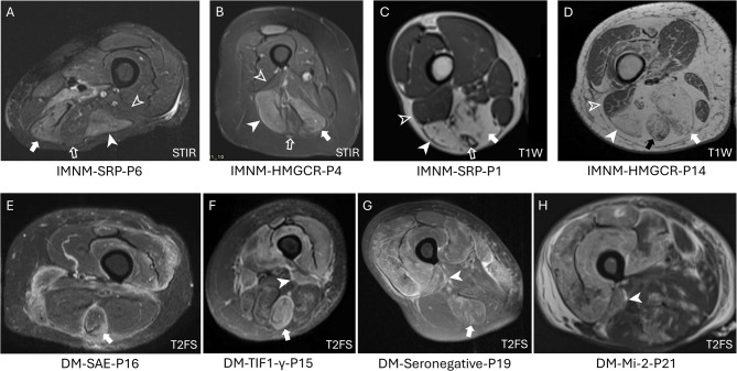
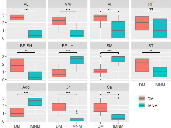
Results: The most affected muscles in IMNM were the semimembranosus (3.0 [2.7-3.0] {median [IQR]}), biceps femoris-long head (BF-LH) (2.7 [2.0-3.0]), and adductors (2.5 [2.0-3.0]). In DM, the most affected muscles were the vastus lateralis (2.7 [2.3-3.0]), vastus intermedius (2.9 [2.2-3.0]), vastus medialis (2.3 [1.7-2.7]), semitendinosus (2.2 [1.0-2.7]), rectus femoris (RF) (2.0 [1.0-2.8]), biceps femoris-short head (BF-SH) (1.9 [1.0-2.7]), gracilis, and sartorius. Intergroup statistical difference of scores was significant (p < 0.01) for 10/11 thigh muscles excluding the RF (p = 0.19), supporting an inverse relationship of muscle involvement for DM and IMNM. The secondary comparative analysis of only muscle edema scores was significant (p < 0.05) for the same 10/11 muscles with a consistent direction for all comparisons.
Conclusion: DM and IMNM affect disparate thigh muscles on MRI. DM preferentially affects the anterior thigh, semitendinosus and BF-SH in the posterior thigh, and gracilis in the medial thigh, whereas IMNM preferentially affects the posterior thigh (semimembranosus and BF-LH) and adductors in the medial thigh.

 求助内容:
求助内容: 应助结果提醒方式:


