{"title":"术前使用肿瘤内和肿瘤周围栖息地成像预测WHO/ISUP分级:多中心研究","authors":"Zhihui Chen, Hongqing Zhu, Hongmin Shu, Jianbo Zhang, Kangchen Gu, Wenjun Yao","doi":"10.1186/s40644-025-00875-z","DOIUrl":null,"url":null,"abstract":"<p><strong>Objectives: </strong>The World Health Organization/International Society of Urological Pathology (WHO/ISUP) grading of clear cell renal cell carcinoma (ccRCC) is crucial for prognosis and treatment planning. This study aims to predict the grade using intratumoral and peritumoral subregional CT radiomics analysis for better clinical interventions.</p><p><strong>Methods: </strong>Data from two hospitals included 513 ccRCC patients, who were divided into training (70%), validation (30%), and an external validation set (testing) of 67 patients. Using ITK-SNAP, two radiologists annotated tumor regions of interest (ROI) and extended surrounding areas by 1 mm, 3 mm, and 5 mm. The K-means clustering algorithm divided the tumor region into three sub-regions, and the Least Absolute Shrinkage and Selection Operator (LASSO) regression identified the most predictive features. Various machine learning models were established, including radiomics models, peritumoral radiomics models, models based on intratumoral heterogeneity (ITH) score, clinical models, and comprehensive models. Predictive ability was evaluated using receiver operating characteristic (ROC) curves, area under the curve (AUC) values, DeLong tests, calibration curves, and decision curves.</p><p><strong>Results: </strong>The combined model showed strong predictive power with an AUC of 0.852 (95% CI: 0.725-0.979) on the test data, outperforming individual models. The ITH score model was highly precise, with AUCs of 0.891 (95% CI: 0.854-0.927) in training, 0.877 (95% CI: 0.814-0.941) in validation, and 0.847 (95% CI: 0.725-0.969) in testing, proving its superior predictive ability across datasets.</p><p><strong>Conclusion: </strong>A comprehensive model combining Habitat, Peri1mm, and salient clinical features was significantly more accurate in predicting ccRCC pathologic grading.</p><p><strong>Key points: </strong>Question: Characterize tumor heterogeneity to non-invasively predict WHO/ISUP pathological grading preoperatively.</p><p><strong>Findings: </strong>An integrated model combining subregion characterization, peritumoral characteristics, and clinical features can predict ccRCC grade preoperatively.</p><p><strong>Clinical relevance: </strong>Subregion tumor characterization outperforms the single-entity approach. The integrated model, compared with the radiomics model, boosts grading and prognostic accuracy for more targeted clinical actions.</p>","PeriodicalId":9548,"journal":{"name":"Cancer Imaging","volume":"25 1","pages":"59"},"PeriodicalIF":3.5000,"publicationDate":"2025-05-03","publicationTypes":"Journal Article","fieldsOfStudy":null,"isOpenAccess":false,"openAccessPdf":"https://www.ncbi.nlm.nih.gov/pmc/articles/PMC12049773/pdf/","citationCount":"0","resultStr":"{\"title\":\"Preoperative prediction of WHO/ISUP grade of ccRCC using intratumoral and peritumoral habitat imaging: multicenter study.\",\"authors\":\"Zhihui Chen, Hongqing Zhu, Hongmin Shu, Jianbo Zhang, Kangchen Gu, Wenjun Yao\",\"doi\":\"10.1186/s40644-025-00875-z\",\"DOIUrl\":null,\"url\":null,\"abstract\":\"<p><strong>Objectives: </strong>The World Health Organization/International Society of Urological Pathology (WHO/ISUP) grading of clear cell renal cell carcinoma (ccRCC) is crucial for prognosis and treatment planning. This study aims to predict the grade using intratumoral and peritumoral subregional CT radiomics analysis for better clinical interventions.</p><p><strong>Methods: </strong>Data from two hospitals included 513 ccRCC patients, who were divided into training (70%), validation (30%), and an external validation set (testing) of 67 patients. Using ITK-SNAP, two radiologists annotated tumor regions of interest (ROI) and extended surrounding areas by 1 mm, 3 mm, and 5 mm. The K-means clustering algorithm divided the tumor region into three sub-regions, and the Least Absolute Shrinkage and Selection Operator (LASSO) regression identified the most predictive features. Various machine learning models were established, including radiomics models, peritumoral radiomics models, models based on intratumoral heterogeneity (ITH) score, clinical models, and comprehensive models. Predictive ability was evaluated using receiver operating characteristic (ROC) curves, area under the curve (AUC) values, DeLong tests, calibration curves, and decision curves.</p><p><strong>Results: </strong>The combined model showed strong predictive power with an AUC of 0.852 (95% CI: 0.725-0.979) on the test data, outperforming individual models. The ITH score model was highly precise, with AUCs of 0.891 (95% CI: 0.854-0.927) in training, 0.877 (95% CI: 0.814-0.941) in validation, and 0.847 (95% CI: 0.725-0.969) in testing, proving its superior predictive ability across datasets.</p><p><strong>Conclusion: </strong>A comprehensive model combining Habitat, Peri1mm, and salient clinical features was significantly more accurate in predicting ccRCC pathologic grading.</p><p><strong>Key points: </strong>Question: Characterize tumor heterogeneity to non-invasively predict WHO/ISUP pathological grading preoperatively.</p><p><strong>Findings: </strong>An integrated model combining subregion characterization, peritumoral characteristics, and clinical features can predict ccRCC grade preoperatively.</p><p><strong>Clinical relevance: </strong>Subregion tumor characterization outperforms the single-entity approach. The integrated model, compared with the radiomics model, boosts grading and prognostic accuracy for more targeted clinical actions.</p>\",\"PeriodicalId\":9548,\"journal\":{\"name\":\"Cancer Imaging\",\"volume\":\"25 1\",\"pages\":\"59\"},\"PeriodicalIF\":3.5000,\"publicationDate\":\"2025-05-03\",\"publicationTypes\":\"Journal Article\",\"fieldsOfStudy\":null,\"isOpenAccess\":false,\"openAccessPdf\":\"https://www.ncbi.nlm.nih.gov/pmc/articles/PMC12049773/pdf/\",\"citationCount\":\"0\",\"resultStr\":null,\"platform\":\"Semanticscholar\",\"paperid\":null,\"PeriodicalName\":\"Cancer Imaging\",\"FirstCategoryId\":\"3\",\"ListUrlMain\":\"https://doi.org/10.1186/s40644-025-00875-z\",\"RegionNum\":2,\"RegionCategory\":\"医学\",\"ArticlePicture\":[],\"TitleCN\":null,\"AbstractTextCN\":null,\"PMCID\":null,\"EPubDate\":\"\",\"PubModel\":\"\",\"JCR\":\"Q2\",\"JCRName\":\"ONCOLOGY\",\"Score\":null,\"Total\":0}","platform":"Semanticscholar","paperid":null,"PeriodicalName":"Cancer Imaging","FirstCategoryId":"3","ListUrlMain":"https://doi.org/10.1186/s40644-025-00875-z","RegionNum":2,"RegionCategory":"医学","ArticlePicture":[],"TitleCN":null,"AbstractTextCN":null,"PMCID":null,"EPubDate":"","PubModel":"","JCR":"Q2","JCRName":"ONCOLOGY","Score":null,"Total":0}
Preoperative prediction of WHO/ISUP grade of ccRCC using intratumoral and peritumoral habitat imaging: multicenter study.
Objectives: The World Health Organization/International Society of Urological Pathology (WHO/ISUP) grading of clear cell renal cell carcinoma (ccRCC) is crucial for prognosis and treatment planning. This study aims to predict the grade using intratumoral and peritumoral subregional CT radiomics analysis for better clinical interventions.
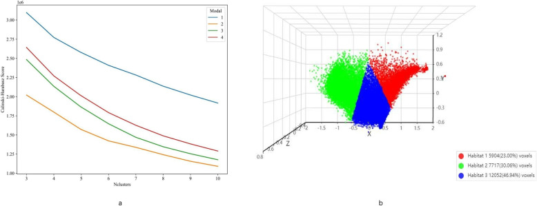
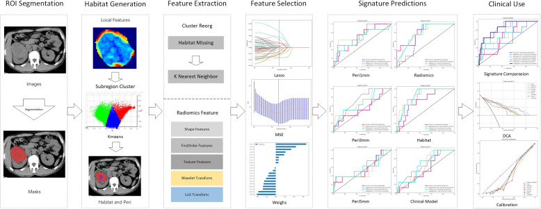
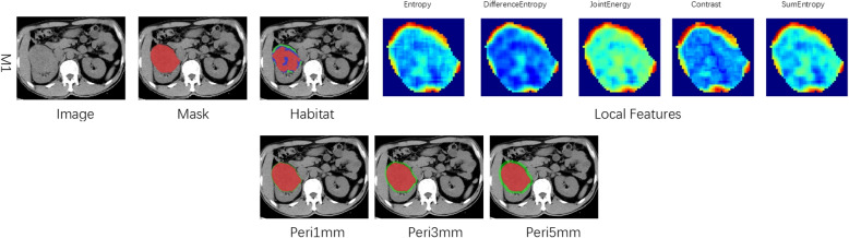
Methods: Data from two hospitals included 513 ccRCC patients, who were divided into training (70%), validation (30%), and an external validation set (testing) of 67 patients. Using ITK-SNAP, two radiologists annotated tumor regions of interest (ROI) and extended surrounding areas by 1 mm, 3 mm, and 5 mm. The K-means clustering algorithm divided the tumor region into three sub-regions, and the Least Absolute Shrinkage and Selection Operator (LASSO) regression identified the most predictive features. Various machine learning models were established, including radiomics models, peritumoral radiomics models, models based on intratumoral heterogeneity (ITH) score, clinical models, and comprehensive models. Predictive ability was evaluated using receiver operating characteristic (ROC) curves, area under the curve (AUC) values, DeLong tests, calibration curves, and decision curves.
Results: The combined model showed strong predictive power with an AUC of 0.852 (95% CI: 0.725-0.979) on the test data, outperforming individual models. The ITH score model was highly precise, with AUCs of 0.891 (95% CI: 0.854-0.927) in training, 0.877 (95% CI: 0.814-0.941) in validation, and 0.847 (95% CI: 0.725-0.969) in testing, proving its superior predictive ability across datasets.
Conclusion: A comprehensive model combining Habitat, Peri1mm, and salient clinical features was significantly more accurate in predicting ccRCC pathologic grading.
Findings: An integrated model combining subregion characterization, peritumoral characteristics, and clinical features can predict ccRCC grade preoperatively.
Clinical relevance: Subregion tumor characterization outperforms the single-entity approach. The integrated model, compared with the radiomics model, boosts grading and prognostic accuracy for more targeted clinical actions.
Cancer ImagingONCOLOGY-RADIOLOGY, NUCLEAR MEDICINE & MEDICAL IMAGING
CiteScore
7.00
自引率
0.00%
发文量
66
审稿时长
>12 weeks
期刊介绍:
Cancer Imaging is an open access, peer-reviewed journal publishing original articles, reviews and editorials written by expert international radiologists working in oncology.
The journal encompasses CT, MR, PET, ultrasound, radionuclide and multimodal imaging in all kinds of malignant tumours, plus new developments, techniques and innovations. Topics of interest include:
Breast Imaging
Chest
Complications of treatment
Ear, Nose & Throat
Gastrointestinal
Hepatobiliary & Pancreatic
Imaging biomarkers
Interventional
Lymphoma
Measurement of tumour response
Molecular functional imaging
Musculoskeletal
Neuro oncology
Nuclear Medicine
Paediatric.

 求助内容:
求助内容: 应助结果提醒方式:


