{"title":"利用深度渐进式学习重建算法增强不同体重指数的18F-FDG PET图像质量和病变诊断性能。","authors":"Zhihao Chen, Hongxing Yang, Ming Qi, Wen Chen, Fei Liu, Shaoli Song, Jianping Zhang","doi":"10.1186/s40644-025-00877-x","DOIUrl":null,"url":null,"abstract":"<p><strong>Background: </strong>As body mass index (BMI) increases, the quality of 2-deoxy-2-[fluorine-18]fluoro-D-glucose (<sup>18</sup>F-FDG) positron emission tomography (PET) images reconstructed with ordered subset expectation maximization (OSEM) declines, negatively impacting lesion diagnostics. It is crucial to identify methods that ensure consistent diagnostic accuracy and maintain image quality. Deep progressive learning (DPL) algorithm, an Artificial Intelligence(AI)-based PET reconstruction technique, offers a promising solution.</p><p><strong>Methods: </strong>150 patients underwent <sup>18</sup>F-FDG PET/CT scans and were categorized by BMI into underweight, normal, and overweight groups. PET images were reconstructed using both OSEM and DPL and their image quality was assessed both visually and quantitatively. Visual assessment employed a 5-point Likert scale to evaluate overall score, image sharpness, image noise, and diagnostic confidence. Quantitative assessment parameters included the background liver image-uniformity-index ([Formula: see text]) and signal-to-noise ratio ([Formula: see text]). Additionally, 466 identifiable lesions were categorized by size: sub-centimeter and larger. We compared maximum standard uptake value ([Formula: see text]), signal-to-background ratio ([Formula: see text]), [Formula: see text], contrast-to-background ratio ([Formula: see text]), and contrast-to-noise ratio ([Formula: see text]) of these lesions to evaluate the diagnostic performance of the DPL and OSEM algorithms across different lesion sizes and BMI categories.</p><p><strong>Results: </strong>DPL produced superior PET image quality compared to OSEM across all BMI groups. The visual quality of DPL showed a slight decline with increasing BMI, while OSEM exhibited a more significant decline. DPL maintained a stable [Formula: see text] across BMI increases, whereas OSEM exhibited increased noise. In the DPL group, quantitative image quality for overweight patients matched that of normal patients with minimal variance from underweight patients. In contrast, OSEM demonstrated significant declines in quantitative image quality with rising BMI. DPL yielded significantly higher contrast ([Formula: see text], [Formula: see text],[Formula: see text]) and [Formula: see text] than OSEM for all lesions across all BMI categories.</p><p><strong>Conclusion: </strong>DPL consistently provided superior image quality and lesion diagnostic performance compared to OSEM across all BMI categories in <sup>18</sup>F-FDG PET/CT scans. Therefore, we recommend using the DPL algorithm for <sup>18</sup>F-FDG PET/CT image reconstruction in all BMI patients.</p>","PeriodicalId":9548,"journal":{"name":"Cancer Imaging","volume":"25 1","pages":"58"},"PeriodicalIF":3.5000,"publicationDate":"2025-05-01","publicationTypes":"Journal Article","fieldsOfStudy":null,"isOpenAccess":false,"openAccessPdf":"https://www.ncbi.nlm.nih.gov/pmc/articles/PMC12044768/pdf/","citationCount":"0","resultStr":"{\"title\":\"Enhancing <sup>18</sup>F-FDG PET image quality and lesion diagnostic performance across different body mass index using the deep progressive learning reconstruction algorithm.\",\"authors\":\"Zhihao Chen, Hongxing Yang, Ming Qi, Wen Chen, Fei Liu, Shaoli Song, Jianping Zhang\",\"doi\":\"10.1186/s40644-025-00877-x\",\"DOIUrl\":null,\"url\":null,\"abstract\":\"<p><strong>Background: </strong>As body mass index (BMI) increases, the quality of 2-deoxy-2-[fluorine-18]fluoro-D-glucose (<sup>18</sup>F-FDG) positron emission tomography (PET) images reconstructed with ordered subset expectation maximization (OSEM) declines, negatively impacting lesion diagnostics. It is crucial to identify methods that ensure consistent diagnostic accuracy and maintain image quality. Deep progressive learning (DPL) algorithm, an Artificial Intelligence(AI)-based PET reconstruction technique, offers a promising solution.</p><p><strong>Methods: </strong>150 patients underwent <sup>18</sup>F-FDG PET/CT scans and were categorized by BMI into underweight, normal, and overweight groups. PET images were reconstructed using both OSEM and DPL and their image quality was assessed both visually and quantitatively. Visual assessment employed a 5-point Likert scale to evaluate overall score, image sharpness, image noise, and diagnostic confidence. Quantitative assessment parameters included the background liver image-uniformity-index ([Formula: see text]) and signal-to-noise ratio ([Formula: see text]). Additionally, 466 identifiable lesions were categorized by size: sub-centimeter and larger. We compared maximum standard uptake value ([Formula: see text]), signal-to-background ratio ([Formula: see text]), [Formula: see text], contrast-to-background ratio ([Formula: see text]), and contrast-to-noise ratio ([Formula: see text]) of these lesions to evaluate the diagnostic performance of the DPL and OSEM algorithms across different lesion sizes and BMI categories.</p><p><strong>Results: </strong>DPL produced superior PET image quality compared to OSEM across all BMI groups. The visual quality of DPL showed a slight decline with increasing BMI, while OSEM exhibited a more significant decline. DPL maintained a stable [Formula: see text] across BMI increases, whereas OSEM exhibited increased noise. In the DPL group, quantitative image quality for overweight patients matched that of normal patients with minimal variance from underweight patients. In contrast, OSEM demonstrated significant declines in quantitative image quality with rising BMI. DPL yielded significantly higher contrast ([Formula: see text], [Formula: see text],[Formula: see text]) and [Formula: see text] than OSEM for all lesions across all BMI categories.</p><p><strong>Conclusion: </strong>DPL consistently provided superior image quality and lesion diagnostic performance compared to OSEM across all BMI categories in <sup>18</sup>F-FDG PET/CT scans. Therefore, we recommend using the DPL algorithm for <sup>18</sup>F-FDG PET/CT image reconstruction in all BMI patients.</p>\",\"PeriodicalId\":9548,\"journal\":{\"name\":\"Cancer Imaging\",\"volume\":\"25 1\",\"pages\":\"58\"},\"PeriodicalIF\":3.5000,\"publicationDate\":\"2025-05-01\",\"publicationTypes\":\"Journal Article\",\"fieldsOfStudy\":null,\"isOpenAccess\":false,\"openAccessPdf\":\"https://www.ncbi.nlm.nih.gov/pmc/articles/PMC12044768/pdf/\",\"citationCount\":\"0\",\"resultStr\":null,\"platform\":\"Semanticscholar\",\"paperid\":null,\"PeriodicalName\":\"Cancer Imaging\",\"FirstCategoryId\":\"3\",\"ListUrlMain\":\"https://doi.org/10.1186/s40644-025-00877-x\",\"RegionNum\":2,\"RegionCategory\":\"医学\",\"ArticlePicture\":[],\"TitleCN\":null,\"AbstractTextCN\":null,\"PMCID\":null,\"EPubDate\":\"\",\"PubModel\":\"\",\"JCR\":\"Q2\",\"JCRName\":\"ONCOLOGY\",\"Score\":null,\"Total\":0}","platform":"Semanticscholar","paperid":null,"PeriodicalName":"Cancer Imaging","FirstCategoryId":"3","ListUrlMain":"https://doi.org/10.1186/s40644-025-00877-x","RegionNum":2,"RegionCategory":"医学","ArticlePicture":[],"TitleCN":null,"AbstractTextCN":null,"PMCID":null,"EPubDate":"","PubModel":"","JCR":"Q2","JCRName":"ONCOLOGY","Score":null,"Total":0}
Enhancing 18F-FDG PET image quality and lesion diagnostic performance across different body mass index using the deep progressive learning reconstruction algorithm.
Background: As body mass index (BMI) increases, the quality of 2-deoxy-2-[fluorine-18]fluoro-D-glucose (18F-FDG) positron emission tomography (PET) images reconstructed with ordered subset expectation maximization (OSEM) declines, negatively impacting lesion diagnostics. It is crucial to identify methods that ensure consistent diagnostic accuracy and maintain image quality. Deep progressive learning (DPL) algorithm, an Artificial Intelligence(AI)-based PET reconstruction technique, offers a promising solution.
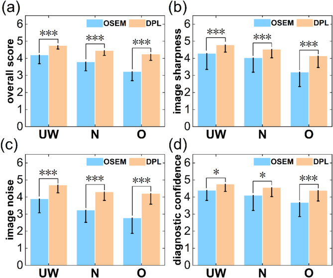
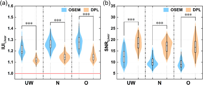
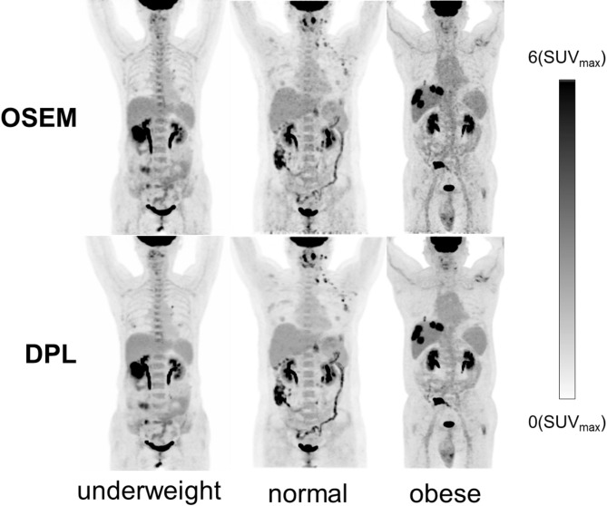
Methods: 150 patients underwent 18F-FDG PET/CT scans and were categorized by BMI into underweight, normal, and overweight groups. PET images were reconstructed using both OSEM and DPL and their image quality was assessed both visually and quantitatively. Visual assessment employed a 5-point Likert scale to evaluate overall score, image sharpness, image noise, and diagnostic confidence. Quantitative assessment parameters included the background liver image-uniformity-index ([Formula: see text]) and signal-to-noise ratio ([Formula: see text]). Additionally, 466 identifiable lesions were categorized by size: sub-centimeter and larger. We compared maximum standard uptake value ([Formula: see text]), signal-to-background ratio ([Formula: see text]), [Formula: see text], contrast-to-background ratio ([Formula: see text]), and contrast-to-noise ratio ([Formula: see text]) of these lesions to evaluate the diagnostic performance of the DPL and OSEM algorithms across different lesion sizes and BMI categories.
Results: DPL produced superior PET image quality compared to OSEM across all BMI groups. The visual quality of DPL showed a slight decline with increasing BMI, while OSEM exhibited a more significant decline. DPL maintained a stable [Formula: see text] across BMI increases, whereas OSEM exhibited increased noise. In the DPL group, quantitative image quality for overweight patients matched that of normal patients with minimal variance from underweight patients. In contrast, OSEM demonstrated significant declines in quantitative image quality with rising BMI. DPL yielded significantly higher contrast ([Formula: see text], [Formula: see text],[Formula: see text]) and [Formula: see text] than OSEM for all lesions across all BMI categories.
Conclusion: DPL consistently provided superior image quality and lesion diagnostic performance compared to OSEM across all BMI categories in 18F-FDG PET/CT scans. Therefore, we recommend using the DPL algorithm for 18F-FDG PET/CT image reconstruction in all BMI patients.
Cancer ImagingONCOLOGY-RADIOLOGY, NUCLEAR MEDICINE & MEDICAL IMAGING
CiteScore
7.00
自引率
0.00%
发文量
66
审稿时长
>12 weeks
期刊介绍:
Cancer Imaging is an open access, peer-reviewed journal publishing original articles, reviews and editorials written by expert international radiologists working in oncology.
The journal encompasses CT, MR, PET, ultrasound, radionuclide and multimodal imaging in all kinds of malignant tumours, plus new developments, techniques and innovations. Topics of interest include:
Breast Imaging
Chest
Complications of treatment
Ear, Nose & Throat
Gastrointestinal
Hepatobiliary & Pancreatic
Imaging biomarkers
Interventional
Lymphoma
Measurement of tumour response
Molecular functional imaging
Musculoskeletal
Neuro oncology
Nuclear Medicine
Paediatric.

 求助内容:
求助内容: 应助结果提醒方式:


