{"title":"光学相干断层扫描血管造影显示视网膜中央凹保留分支动脉闭塞:2例报告。","authors":"Benjamin R Lin, Philip J Rosenfeld, Harry W Flynn","doi":"10.1159/000543742","DOIUrl":null,"url":null,"abstract":"<p><strong>Introduction: </strong>Central retinal artery occlusions and branch retinal artery occlusions (BRAOs) are ophthalmic emergencies that require workups for systemic risk factors. In the acute setting, BRAOs present with retinal whitening in a sectoral pattern on exam as well as hyperreflectivity and thickening of the inner retinal layers on optical coherence tomography (OCT). In the subacute to chronic phase, the retinal whitening dissipates, which may confound the diagnosis of remote arterial occlusions if there is no clearly visible plaque.</p><p><strong>Case presentations: </strong>A 66-year-old male presented with 20/25 visual acuity (VA) and an inferior visual field defect in the right eye, and a 69-year-old male presented with 20/60 VA and a superior visual field defect in the left eye. Exams of both patients showed ischemic retinal whitening with visible Hollenhorst plaques in the affected eyes. OCT demonstrated inner retinal edema. At follow-up, wide-field OCT angiography (OCTA) showed persistent capillary dropout following the same initial vascular distribution but sparing the fovea and papillomacular bundle. VAs at the most recent follow-up visits were 20/30 and 20/20, respectively.</p><p><strong>Conclusion: </strong>These cases demonstrate the utility of wide-field OCTA in characterizing areas of capillary nonperfusion that can persist for years after the initial ischemic event. Additionally, patients with macula-involving BRAOs can have good VA outcomes if the fovea is spared.</p>","PeriodicalId":9635,"journal":{"name":"Case Reports in Ophthalmology","volume":"16 1","pages":"274-280"},"PeriodicalIF":0.6000,"publicationDate":"2025-03-12","publicationTypes":"Journal Article","fieldsOfStudy":null,"isOpenAccess":false,"openAccessPdf":"https://www.ncbi.nlm.nih.gov/pmc/articles/PMC12005688/pdf/","citationCount":"0","resultStr":"{\"title\":\"Fovea Sparing Branch Retinal Artery Occlusions Imaged with Optical Coherence Tomography Angiography: Two Case Reports.\",\"authors\":\"Benjamin R Lin, Philip J Rosenfeld, Harry W Flynn\",\"doi\":\"10.1159/000543742\",\"DOIUrl\":null,\"url\":null,\"abstract\":\"<p><strong>Introduction: </strong>Central retinal artery occlusions and branch retinal artery occlusions (BRAOs) are ophthalmic emergencies that require workups for systemic risk factors. In the acute setting, BRAOs present with retinal whitening in a sectoral pattern on exam as well as hyperreflectivity and thickening of the inner retinal layers on optical coherence tomography (OCT). In the subacute to chronic phase, the retinal whitening dissipates, which may confound the diagnosis of remote arterial occlusions if there is no clearly visible plaque.</p><p><strong>Case presentations: </strong>A 66-year-old male presented with 20/25 visual acuity (VA) and an inferior visual field defect in the right eye, and a 69-year-old male presented with 20/60 VA and a superior visual field defect in the left eye. Exams of both patients showed ischemic retinal whitening with visible Hollenhorst plaques in the affected eyes. OCT demonstrated inner retinal edema. At follow-up, wide-field OCT angiography (OCTA) showed persistent capillary dropout following the same initial vascular distribution but sparing the fovea and papillomacular bundle. VAs at the most recent follow-up visits were 20/30 and 20/20, respectively.</p><p><strong>Conclusion: </strong>These cases demonstrate the utility of wide-field OCTA in characterizing areas of capillary nonperfusion that can persist for years after the initial ischemic event. Additionally, patients with macula-involving BRAOs can have good VA outcomes if the fovea is spared.</p>\",\"PeriodicalId\":9635,\"journal\":{\"name\":\"Case Reports in Ophthalmology\",\"volume\":\"16 1\",\"pages\":\"274-280\"},\"PeriodicalIF\":0.6000,\"publicationDate\":\"2025-03-12\",\"publicationTypes\":\"Journal Article\",\"fieldsOfStudy\":null,\"isOpenAccess\":false,\"openAccessPdf\":\"https://www.ncbi.nlm.nih.gov/pmc/articles/PMC12005688/pdf/\",\"citationCount\":\"0\",\"resultStr\":null,\"platform\":\"Semanticscholar\",\"paperid\":null,\"PeriodicalName\":\"Case Reports in Ophthalmology\",\"FirstCategoryId\":\"1085\",\"ListUrlMain\":\"https://doi.org/10.1159/000543742\",\"RegionNum\":0,\"RegionCategory\":null,\"ArticlePicture\":[],\"TitleCN\":null,\"AbstractTextCN\":null,\"PMCID\":null,\"EPubDate\":\"2025/1/1 0:00:00\",\"PubModel\":\"eCollection\",\"JCR\":\"Q4\",\"JCRName\":\"OPHTHALMOLOGY\",\"Score\":null,\"Total\":0}","platform":"Semanticscholar","paperid":null,"PeriodicalName":"Case Reports in Ophthalmology","FirstCategoryId":"1085","ListUrlMain":"https://doi.org/10.1159/000543742","RegionNum":0,"RegionCategory":null,"ArticlePicture":[],"TitleCN":null,"AbstractTextCN":null,"PMCID":null,"EPubDate":"2025/1/1 0:00:00","PubModel":"eCollection","JCR":"Q4","JCRName":"OPHTHALMOLOGY","Score":null,"Total":0}
Fovea Sparing Branch Retinal Artery Occlusions Imaged with Optical Coherence Tomography Angiography: Two Case Reports.
Introduction: Central retinal artery occlusions and branch retinal artery occlusions (BRAOs) are ophthalmic emergencies that require workups for systemic risk factors. In the acute setting, BRAOs present with retinal whitening in a sectoral pattern on exam as well as hyperreflectivity and thickening of the inner retinal layers on optical coherence tomography (OCT). In the subacute to chronic phase, the retinal whitening dissipates, which may confound the diagnosis of remote arterial occlusions if there is no clearly visible plaque.
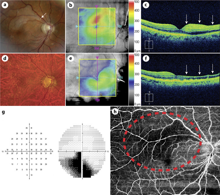
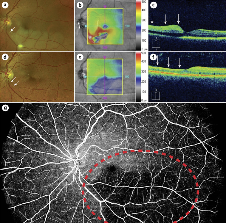
Case presentations: A 66-year-old male presented with 20/25 visual acuity (VA) and an inferior visual field defect in the right eye, and a 69-year-old male presented with 20/60 VA and a superior visual field defect in the left eye. Exams of both patients showed ischemic retinal whitening with visible Hollenhorst plaques in the affected eyes. OCT demonstrated inner retinal edema. At follow-up, wide-field OCT angiography (OCTA) showed persistent capillary dropout following the same initial vascular distribution but sparing the fovea and papillomacular bundle. VAs at the most recent follow-up visits were 20/30 and 20/20, respectively.
Conclusion: These cases demonstrate the utility of wide-field OCTA in characterizing areas of capillary nonperfusion that can persist for years after the initial ischemic event. Additionally, patients with macula-involving BRAOs can have good VA outcomes if the fovea is spared.
期刊介绍:
This peer-reviewed online-only journal publishes original case reports covering the entire spectrum of ophthalmology, including prevention, diagnosis, treatment, toxicities of therapy, supportive care, quality-of-life, and survivorship issues. The submission of negative results is strongly encouraged. The journal will also accept case reports dealing with the use of novel technologies, both in the arena of diagnosis and treatment. Supplementary material is welcomed. The intent of the journal is to provide clinicians and researchers with a tool to disseminate their personal experiences to a wider public as well as to review interesting cases encountered by colleagues all over the world. Universally used terms can be searched across the entire growing collection of case reports, further facilitating the retrieval of specific information. Following the open access principle, the entire contents can be retrieved at no charge, guaranteeing easy access to this valuable source of anecdotal information at all times.

 求助内容:
求助内容: 应助结果提醒方式:


