Yuqi Yan, Yuanzhen Liu, Yao Wang, Tian Jiang, Jiayu Xie, Yahan Zhou, Xin Liu, Meiying Yan, Qiuqing Zheng, Haifei Xu, Jinxiao Chen, Lin Sui, Chen Chen, RongRong Ru, Kai Wang, Anli Zhao, Shiyan Li, Ying Zhu, Yang Zhang, Vicky Yang Wang, Dong Xu
{"title":"基于超声图像深度学习的乳腺叶状肿瘤分级诊断:一项回顾性多中心研究。","authors":"Yuqi Yan, Yuanzhen Liu, Yao Wang, Tian Jiang, Jiayu Xie, Yahan Zhou, Xin Liu, Meiying Yan, Qiuqing Zheng, Haifei Xu, Jinxiao Chen, Lin Sui, Chen Chen, RongRong Ru, Kai Wang, Anli Zhao, Shiyan Li, Ying Zhu, Yang Zhang, Vicky Yang Wang, Dong Xu","doi":"10.1186/s40644-025-00879-9","DOIUrl":null,"url":null,"abstract":"<p><strong>Objective: </strong>Phyllodes tumors (PTs) are rare breast tumors with high recurrence rates, current methods relying on post-resection pathology often delay detection and require further surgery. We propose a deep-learning-based Phyllodes Tumors Hierarchical Diagnosis Model (PTs-HDM) for preoperative identification and grading.</p><p><strong>Methods: </strong>Ultrasound images from five hospitals were retrospectively collected, with all patients having undergone surgical pathological confirmation of either PTs or fibroadenomas (FAs). PTs-HDM follows a two-stage classification: first distinguishing PTs from FAs, then grading PTs into benign or borderline/malignant. Model performance metrics including AUC and accuracy were quantitatively evaluated. A comparative analysis was conducted between the algorithm's diagnostic capabilities and those of radiologists with varying clinical experience within an external validation cohort. Through the provision of PTs-HDM's automated classification outputs and associated thermal activation mapping guidance, we systematically assessed the enhancement in radiologists' diagnostic concordance and classification accuracy.</p><p><strong>Results: </strong>A total of 712 patients were included. On the external test set, PTs-HDM achieved an AUC of 0.883, accuracy of 87.3% for PT vs. FA classification. Subgroup analysis showed high accuracy for tumors < 2 cm (90.9%). In hierarchical classification, the model obtained an AUC of 0.856 and accuracy of 80.9%. Radiologists' performance improved with PTs-HDM assistance, with binary classification accuracy increasing from 82.7%, 67.7%, and 64.2-87.6%, 76.6%, and 82.1% for senior, attending, and resident radiologists, respectively. Their hierarchical classification AUCs improved from 0.566 to 0.827 to 0.725-0.837. PTs-HDM also enhanced inter-radiologist consistency, increasing Kappa values from - 0.05 to 0.41 to 0.12 to 0.65, and the intraclass correlation coefficient from 0.19 to 0.45.</p><p><strong>Conclusion: </strong>PTs-HDM shows strong diagnostic performance, especially for small lesions, and improves radiologists' accuracy across all experience levels, bridging diagnostic gaps and providing reliable support for PTs' hierarchical diagnosis.</p>","PeriodicalId":9548,"journal":{"name":"Cancer Imaging","volume":"25 1","pages":"61"},"PeriodicalIF":3.5000,"publicationDate":"2025-05-08","publicationTypes":"Journal Article","fieldsOfStudy":null,"isOpenAccess":false,"openAccessPdf":"https://www.ncbi.nlm.nih.gov/pmc/articles/PMC12063467/pdf/","citationCount":"0","resultStr":"{\"title\":\"Hierarchical diagnosis of breast phyllodes tumors enabled by deep learning of ultrasound images: a retrospective multi-center study.\",\"authors\":\"Yuqi Yan, Yuanzhen Liu, Yao Wang, Tian Jiang, Jiayu Xie, Yahan Zhou, Xin Liu, Meiying Yan, Qiuqing Zheng, Haifei Xu, Jinxiao Chen, Lin Sui, Chen Chen, RongRong Ru, Kai Wang, Anli Zhao, Shiyan Li, Ying Zhu, Yang Zhang, Vicky Yang Wang, Dong Xu\",\"doi\":\"10.1186/s40644-025-00879-9\",\"DOIUrl\":null,\"url\":null,\"abstract\":\"<p><strong>Objective: </strong>Phyllodes tumors (PTs) are rare breast tumors with high recurrence rates, current methods relying on post-resection pathology often delay detection and require further surgery. We propose a deep-learning-based Phyllodes Tumors Hierarchical Diagnosis Model (PTs-HDM) for preoperative identification and grading.</p><p><strong>Methods: </strong>Ultrasound images from five hospitals were retrospectively collected, with all patients having undergone surgical pathological confirmation of either PTs or fibroadenomas (FAs). PTs-HDM follows a two-stage classification: first distinguishing PTs from FAs, then grading PTs into benign or borderline/malignant. Model performance metrics including AUC and accuracy were quantitatively evaluated. A comparative analysis was conducted between the algorithm's diagnostic capabilities and those of radiologists with varying clinical experience within an external validation cohort. Through the provision of PTs-HDM's automated classification outputs and associated thermal activation mapping guidance, we systematically assessed the enhancement in radiologists' diagnostic concordance and classification accuracy.</p><p><strong>Results: </strong>A total of 712 patients were included. On the external test set, PTs-HDM achieved an AUC of 0.883, accuracy of 87.3% for PT vs. FA classification. Subgroup analysis showed high accuracy for tumors < 2 cm (90.9%). In hierarchical classification, the model obtained an AUC of 0.856 and accuracy of 80.9%. Radiologists' performance improved with PTs-HDM assistance, with binary classification accuracy increasing from 82.7%, 67.7%, and 64.2-87.6%, 76.6%, and 82.1% for senior, attending, and resident radiologists, respectively. Their hierarchical classification AUCs improved from 0.566 to 0.827 to 0.725-0.837. PTs-HDM also enhanced inter-radiologist consistency, increasing Kappa values from - 0.05 to 0.41 to 0.12 to 0.65, and the intraclass correlation coefficient from 0.19 to 0.45.</p><p><strong>Conclusion: </strong>PTs-HDM shows strong diagnostic performance, especially for small lesions, and improves radiologists' accuracy across all experience levels, bridging diagnostic gaps and providing reliable support for PTs' hierarchical diagnosis.</p>\",\"PeriodicalId\":9548,\"journal\":{\"name\":\"Cancer Imaging\",\"volume\":\"25 1\",\"pages\":\"61\"},\"PeriodicalIF\":3.5000,\"publicationDate\":\"2025-05-08\",\"publicationTypes\":\"Journal Article\",\"fieldsOfStudy\":null,\"isOpenAccess\":false,\"openAccessPdf\":\"https://www.ncbi.nlm.nih.gov/pmc/articles/PMC12063467/pdf/\",\"citationCount\":\"0\",\"resultStr\":null,\"platform\":\"Semanticscholar\",\"paperid\":null,\"PeriodicalName\":\"Cancer Imaging\",\"FirstCategoryId\":\"3\",\"ListUrlMain\":\"https://doi.org/10.1186/s40644-025-00879-9\",\"RegionNum\":2,\"RegionCategory\":\"医学\",\"ArticlePicture\":[],\"TitleCN\":null,\"AbstractTextCN\":null,\"PMCID\":null,\"EPubDate\":\"\",\"PubModel\":\"\",\"JCR\":\"Q2\",\"JCRName\":\"ONCOLOGY\",\"Score\":null,\"Total\":0}","platform":"Semanticscholar","paperid":null,"PeriodicalName":"Cancer Imaging","FirstCategoryId":"3","ListUrlMain":"https://doi.org/10.1186/s40644-025-00879-9","RegionNum":2,"RegionCategory":"医学","ArticlePicture":[],"TitleCN":null,"AbstractTextCN":null,"PMCID":null,"EPubDate":"","PubModel":"","JCR":"Q2","JCRName":"ONCOLOGY","Score":null,"Total":0}
Hierarchical diagnosis of breast phyllodes tumors enabled by deep learning of ultrasound images: a retrospective multi-center study.
Objective: Phyllodes tumors (PTs) are rare breast tumors with high recurrence rates, current methods relying on post-resection pathology often delay detection and require further surgery. We propose a deep-learning-based Phyllodes Tumors Hierarchical Diagnosis Model (PTs-HDM) for preoperative identification and grading.
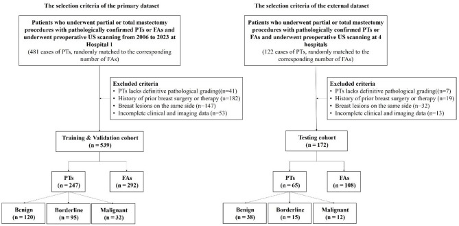
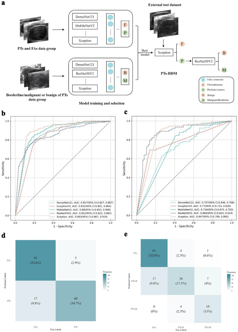
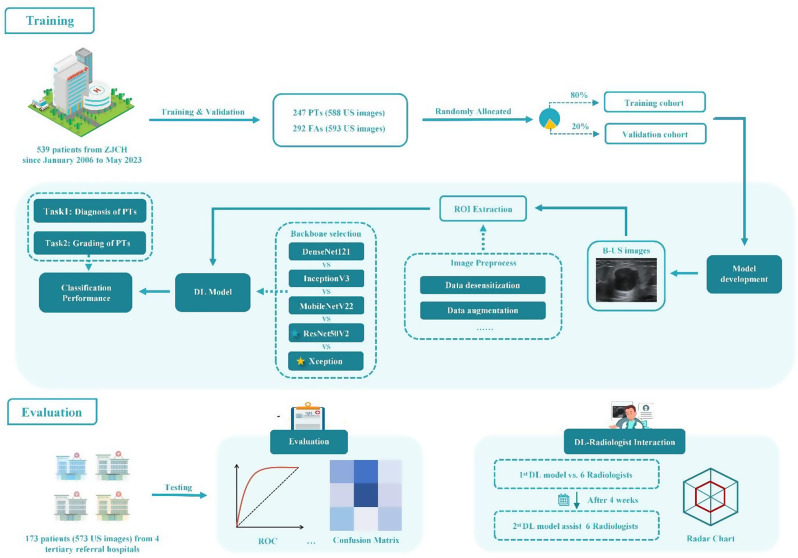
Methods: Ultrasound images from five hospitals were retrospectively collected, with all patients having undergone surgical pathological confirmation of either PTs or fibroadenomas (FAs). PTs-HDM follows a two-stage classification: first distinguishing PTs from FAs, then grading PTs into benign or borderline/malignant. Model performance metrics including AUC and accuracy were quantitatively evaluated. A comparative analysis was conducted between the algorithm's diagnostic capabilities and those of radiologists with varying clinical experience within an external validation cohort. Through the provision of PTs-HDM's automated classification outputs and associated thermal activation mapping guidance, we systematically assessed the enhancement in radiologists' diagnostic concordance and classification accuracy.
Results: A total of 712 patients were included. On the external test set, PTs-HDM achieved an AUC of 0.883, accuracy of 87.3% for PT vs. FA classification. Subgroup analysis showed high accuracy for tumors < 2 cm (90.9%). In hierarchical classification, the model obtained an AUC of 0.856 and accuracy of 80.9%. Radiologists' performance improved with PTs-HDM assistance, with binary classification accuracy increasing from 82.7%, 67.7%, and 64.2-87.6%, 76.6%, and 82.1% for senior, attending, and resident radiologists, respectively. Their hierarchical classification AUCs improved from 0.566 to 0.827 to 0.725-0.837. PTs-HDM also enhanced inter-radiologist consistency, increasing Kappa values from - 0.05 to 0.41 to 0.12 to 0.65, and the intraclass correlation coefficient from 0.19 to 0.45.
Conclusion: PTs-HDM shows strong diagnostic performance, especially for small lesions, and improves radiologists' accuracy across all experience levels, bridging diagnostic gaps and providing reliable support for PTs' hierarchical diagnosis.
Cancer ImagingONCOLOGY-RADIOLOGY, NUCLEAR MEDICINE & MEDICAL IMAGING
CiteScore
7.00
自引率
0.00%
发文量
66
审稿时长
>12 weeks
期刊介绍:
Cancer Imaging is an open access, peer-reviewed journal publishing original articles, reviews and editorials written by expert international radiologists working in oncology.
The journal encompasses CT, MR, PET, ultrasound, radionuclide and multimodal imaging in all kinds of malignant tumours, plus new developments, techniques and innovations. Topics of interest include:
Breast Imaging
Chest
Complications of treatment
Ear, Nose & Throat
Gastrointestinal
Hepatobiliary & Pancreatic
Imaging biomarkers
Interventional
Lymphoma
Measurement of tumour response
Molecular functional imaging
Musculoskeletal
Neuro oncology
Nuclear Medicine
Paediatric.

 求助内容:
求助内容: 应助结果提醒方式:


