Živa Ledinek, Milica Stefanović, Blaž Mavčič, Maja Česen Mazić, Ana Gazikalović, Daja Šekoranja, Simona Miceska, Veronika Kloboves Prevodnik
{"title":"基于FNAB样本的细胞形态学和分子分析诊断小儿骨化性肌炎1例。","authors":"Živa Ledinek, Milica Stefanović, Blaž Mavčič, Maja Česen Mazić, Ana Gazikalović, Daja Šekoranja, Simona Miceska, Veronika Kloboves Prevodnik","doi":"10.1002/dc.25477","DOIUrl":null,"url":null,"abstract":"<p>Myositis ossificans (MO) is a benign soft tissue lesion, characterized by ectopic ossification due to inappropriate fibroblast differentiation, most commonly affecting skeletal muscles. It often occurs in young adults after muscle trauma, predominantly in male patients and very rarely in children. We describe the case of a previously healthy 3-year-old boy who developed a lesion in his deltoid muscle after vaccination against tick-borne encephalitis. During an MRI scan, performed under general anesthesia, fine needle aspiration biopsy (FNAB) and core needle biopsy (CNB) were performed. While the CNB sample resulted in a non-diagnostic finding, the FNAB sample showed cytomorphology consistent with the diagnosis of MO. A molecular analysis performed on the FNAB sample confirmed the presence of <i>COL1A1::USP6</i> fusion, which is considered diagnostic for MO in the appropriate clinical context. The boy was then referred to the orthopedic surgeon. Extracorporeal shock-wave therapy was chosen as the first-line treatment, but as it was too painful, the lesion was surgically removed, and histopathologic evaluation confirmed the diagnosis. Although rare in children, the diagnosis of MO should be considered in soft tissue lesions after trauma. We present the first pediatric case of MO diagnosed by FNAB and propose that FNAB, as a minimally invasive diagnostic procedure, is a suitable diagnostic approach, especially when molecular testing is available to confirm the diagnosis.</p>","PeriodicalId":11349,"journal":{"name":"Diagnostic Cytopathology","volume":"53 7","pages":"E138-E143"},"PeriodicalIF":1.0000,"publicationDate":"2025-04-22","publicationTypes":"Journal Article","fieldsOfStudy":null,"isOpenAccess":false,"openAccessPdf":"https://onlinelibrary.wiley.com/doi/epdf/10.1002/dc.25477","citationCount":"0","resultStr":"{\"title\":\"Diagnosis of Pediatric Myositis Ossificans Based on Cytomorphology and Molecular Analysis From FNAB Sample: A Case Report\",\"authors\":\"Živa Ledinek, Milica Stefanović, Blaž Mavčič, Maja Česen Mazić, Ana Gazikalović, Daja Šekoranja, Simona Miceska, Veronika Kloboves Prevodnik\",\"doi\":\"10.1002/dc.25477\",\"DOIUrl\":null,\"url\":null,\"abstract\":\"<p>Myositis ossificans (MO) is a benign soft tissue lesion, characterized by ectopic ossification due to inappropriate fibroblast differentiation, most commonly affecting skeletal muscles. It often occurs in young adults after muscle trauma, predominantly in male patients and very rarely in children. We describe the case of a previously healthy 3-year-old boy who developed a lesion in his deltoid muscle after vaccination against tick-borne encephalitis. During an MRI scan, performed under general anesthesia, fine needle aspiration biopsy (FNAB) and core needle biopsy (CNB) were performed. While the CNB sample resulted in a non-diagnostic finding, the FNAB sample showed cytomorphology consistent with the diagnosis of MO. A molecular analysis performed on the FNAB sample confirmed the presence of <i>COL1A1::USP6</i> fusion, which is considered diagnostic for MO in the appropriate clinical context. The boy was then referred to the orthopedic surgeon. Extracorporeal shock-wave therapy was chosen as the first-line treatment, but as it was too painful, the lesion was surgically removed, and histopathologic evaluation confirmed the diagnosis. Although rare in children, the diagnosis of MO should be considered in soft tissue lesions after trauma. We present the first pediatric case of MO diagnosed by FNAB and propose that FNAB, as a minimally invasive diagnostic procedure, is a suitable diagnostic approach, especially when molecular testing is available to confirm the diagnosis.</p>\",\"PeriodicalId\":11349,\"journal\":{\"name\":\"Diagnostic Cytopathology\",\"volume\":\"53 7\",\"pages\":\"E138-E143\"},\"PeriodicalIF\":1.0000,\"publicationDate\":\"2025-04-22\",\"publicationTypes\":\"Journal Article\",\"fieldsOfStudy\":null,\"isOpenAccess\":false,\"openAccessPdf\":\"https://onlinelibrary.wiley.com/doi/epdf/10.1002/dc.25477\",\"citationCount\":\"0\",\"resultStr\":null,\"platform\":\"Semanticscholar\",\"paperid\":null,\"PeriodicalName\":\"Diagnostic Cytopathology\",\"FirstCategoryId\":\"3\",\"ListUrlMain\":\"https://onlinelibrary.wiley.com/doi/10.1002/dc.25477\",\"RegionNum\":4,\"RegionCategory\":\"医学\",\"ArticlePicture\":[],\"TitleCN\":null,\"AbstractTextCN\":null,\"PMCID\":null,\"EPubDate\":\"\",\"PubModel\":\"\",\"JCR\":\"Q4\",\"JCRName\":\"MEDICAL LABORATORY TECHNOLOGY\",\"Score\":null,\"Total\":0}","platform":"Semanticscholar","paperid":null,"PeriodicalName":"Diagnostic Cytopathology","FirstCategoryId":"3","ListUrlMain":"https://onlinelibrary.wiley.com/doi/10.1002/dc.25477","RegionNum":4,"RegionCategory":"医学","ArticlePicture":[],"TitleCN":null,"AbstractTextCN":null,"PMCID":null,"EPubDate":"","PubModel":"","JCR":"Q4","JCRName":"MEDICAL LABORATORY TECHNOLOGY","Score":null,"Total":0}
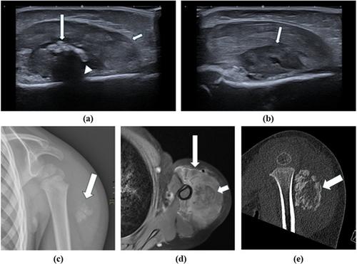
Diagnosis of Pediatric Myositis Ossificans Based on Cytomorphology and Molecular Analysis From FNAB Sample: A Case Report
Myositis ossificans (MO) is a benign soft tissue lesion, characterized by ectopic ossification due to inappropriate fibroblast differentiation, most commonly affecting skeletal muscles. It often occurs in young adults after muscle trauma, predominantly in male patients and very rarely in children. We describe the case of a previously healthy 3-year-old boy who developed a lesion in his deltoid muscle after vaccination against tick-borne encephalitis. During an MRI scan, performed under general anesthesia, fine needle aspiration biopsy (FNAB) and core needle biopsy (CNB) were performed. While the CNB sample resulted in a non-diagnostic finding, the FNAB sample showed cytomorphology consistent with the diagnosis of MO. A molecular analysis performed on the FNAB sample confirmed the presence of COL1A1::USP6 fusion, which is considered diagnostic for MO in the appropriate clinical context. The boy was then referred to the orthopedic surgeon. Extracorporeal shock-wave therapy was chosen as the first-line treatment, but as it was too painful, the lesion was surgically removed, and histopathologic evaluation confirmed the diagnosis. Although rare in children, the diagnosis of MO should be considered in soft tissue lesions after trauma. We present the first pediatric case of MO diagnosed by FNAB and propose that FNAB, as a minimally invasive diagnostic procedure, is a suitable diagnostic approach, especially when molecular testing is available to confirm the diagnosis.
期刊介绍:
Diagnostic Cytopathology is intended to provide a forum for the exchange of information in the field of cytopathology, with special emphasis on the practical, clinical aspects of the discipline. The editors invite original scientific articles, as well as special review articles, feature articles, and letters to the editor, from laboratory professionals engaged in the practice of cytopathology. Manuscripts are accepted for publication on the basis of scientific merit, practical significance, and suitability for publication in a journal dedicated to this discipline. Original articles can be considered only with the understanding that they have never been published before and that they have not been submitted for simultaneous review to another publication.

 求助内容:
求助内容: 应助结果提醒方式:


