{"title":"深入了解平山病:颈椎以外的硬脑膜脱离。","authors":"Seena Vengalil, Vijaykumar Boddu, Karthik Kulanthaivelu, Dipti Baskar, Saraswati Nashi, Nupur Pruthi, Hemant Bhargav, Alok M Uppar, Chandrajit Prasad, Madhulika Kotra, Kiran Polavarapu, Veeramani Preethish-Kumar, Atchayaram Nalini","doi":"10.4103/aian.aian_16_25","DOIUrl":null,"url":null,"abstract":"<p><strong>Background and objectives: </strong>Hirayama disease (HD) is a cervical flexion-induced compressive myelopathy. Typically, there is forward displacement and loss of attachment of dural sac to lamina at the cervical level during neck flexion. However, the extent of the dural detachment (DD) has not been studied carefully. We undertook this study to know the extent of DD in HD.</p><p><strong>Methods: </strong>We conducted a retrospective study of HD patients evaluated from 2015 to 2023. Patients with DD extending beyond the cervical spine were selected, and their clinical and radiological features were studied.</p><p><strong>Results: </strong>One hundred and thirty-two (62.8%) patients were identified to have DD beyond the cervical spine in a cohort of 210 HD patients. The mean age at onset and duration were 18.09 ± 2.3 years (13-26) and 38.63 ± 39.9 months, respectively. Proximo-distal involvement was noted in 50% of patients, while 33% and 17% of patients had isolated distal and proximal involvement, respectively. Wasted legs were observed in three patients. Cord atrophy was present in 96.9% of patients, extending from C5 to C7. Epidural detachment and engorgement of posterior epidural venous plexus were evident in all. DD extended from C2 to D10 vertebral level. The cranial extent of DD was from C2 to C4 in 87% of cases, and the caudal extent was D1-D5 in 84% of cases, extending up to D10 in two cases.</p><p><strong>Conclusions: </strong>The HD spectrum continues to evolve phenotypically and radiologically. The pathophysiological mechanisms and DD extend beyond the cervical spine in a large proportion of patients. This makes it important to cover a longer part of the spine during imaging. This may have implications on the management of patients, particularly those with isolated lower limb involvement.</p>","PeriodicalId":8036,"journal":{"name":"Annals of Indian Academy of Neurology","volume":" ","pages":"574-578"},"PeriodicalIF":1.8000,"publicationDate":"2025-07-01","publicationTypes":"Journal Article","fieldsOfStudy":null,"isOpenAccess":false,"openAccessPdf":"https://www.ncbi.nlm.nih.gov/pmc/articles/PMC12393858/pdf/","citationCount":"0","resultStr":"{\"title\":\"In-Depth Understanding of Hirayama Disease: Dural Detachment Beyond Cervical Spine.\",\"authors\":\"Seena Vengalil, Vijaykumar Boddu, Karthik Kulanthaivelu, Dipti Baskar, Saraswati Nashi, Nupur Pruthi, Hemant Bhargav, Alok M Uppar, Chandrajit Prasad, Madhulika Kotra, Kiran Polavarapu, Veeramani Preethish-Kumar, Atchayaram Nalini\",\"doi\":\"10.4103/aian.aian_16_25\",\"DOIUrl\":null,\"url\":null,\"abstract\":\"<p><strong>Background and objectives: </strong>Hirayama disease (HD) is a cervical flexion-induced compressive myelopathy. Typically, there is forward displacement and loss of attachment of dural sac to lamina at the cervical level during neck flexion. However, the extent of the dural detachment (DD) has not been studied carefully. We undertook this study to know the extent of DD in HD.</p><p><strong>Methods: </strong>We conducted a retrospective study of HD patients evaluated from 2015 to 2023. Patients with DD extending beyond the cervical spine were selected, and their clinical and radiological features were studied.</p><p><strong>Results: </strong>One hundred and thirty-two (62.8%) patients were identified to have DD beyond the cervical spine in a cohort of 210 HD patients. The mean age at onset and duration were 18.09 ± 2.3 years (13-26) and 38.63 ± 39.9 months, respectively. Proximo-distal involvement was noted in 50% of patients, while 33% and 17% of patients had isolated distal and proximal involvement, respectively. Wasted legs were observed in three patients. Cord atrophy was present in 96.9% of patients, extending from C5 to C7. Epidural detachment and engorgement of posterior epidural venous plexus were evident in all. DD extended from C2 to D10 vertebral level. The cranial extent of DD was from C2 to C4 in 87% of cases, and the caudal extent was D1-D5 in 84% of cases, extending up to D10 in two cases.</p><p><strong>Conclusions: </strong>The HD spectrum continues to evolve phenotypically and radiologically. The pathophysiological mechanisms and DD extend beyond the cervical spine in a large proportion of patients. This makes it important to cover a longer part of the spine during imaging. This may have implications on the management of patients, particularly those with isolated lower limb involvement.</p>\",\"PeriodicalId\":8036,\"journal\":{\"name\":\"Annals of Indian Academy of Neurology\",\"volume\":\" \",\"pages\":\"574-578\"},\"PeriodicalIF\":1.8000,\"publicationDate\":\"2025-07-01\",\"publicationTypes\":\"Journal Article\",\"fieldsOfStudy\":null,\"isOpenAccess\":false,\"openAccessPdf\":\"https://www.ncbi.nlm.nih.gov/pmc/articles/PMC12393858/pdf/\",\"citationCount\":\"0\",\"resultStr\":null,\"platform\":\"Semanticscholar\",\"paperid\":null,\"PeriodicalName\":\"Annals of Indian Academy of Neurology\",\"FirstCategoryId\":\"3\",\"ListUrlMain\":\"https://doi.org/10.4103/aian.aian_16_25\",\"RegionNum\":4,\"RegionCategory\":\"医学\",\"ArticlePicture\":[],\"TitleCN\":null,\"AbstractTextCN\":null,\"PMCID\":null,\"EPubDate\":\"2025/4/22 0:00:00\",\"PubModel\":\"Epub\",\"JCR\":\"Q3\",\"JCRName\":\"CLINICAL NEUROLOGY\",\"Score\":null,\"Total\":0}","platform":"Semanticscholar","paperid":null,"PeriodicalName":"Annals of Indian Academy of Neurology","FirstCategoryId":"3","ListUrlMain":"https://doi.org/10.4103/aian.aian_16_25","RegionNum":4,"RegionCategory":"医学","ArticlePicture":[],"TitleCN":null,"AbstractTextCN":null,"PMCID":null,"EPubDate":"2025/4/22 0:00:00","PubModel":"Epub","JCR":"Q3","JCRName":"CLINICAL NEUROLOGY","Score":null,"Total":0}
In-Depth Understanding of Hirayama Disease: Dural Detachment Beyond Cervical Spine.
Background and objectives: Hirayama disease (HD) is a cervical flexion-induced compressive myelopathy. Typically, there is forward displacement and loss of attachment of dural sac to lamina at the cervical level during neck flexion. However, the extent of the dural detachment (DD) has not been studied carefully. We undertook this study to know the extent of DD in HD.
Methods: We conducted a retrospective study of HD patients evaluated from 2015 to 2023. Patients with DD extending beyond the cervical spine were selected, and their clinical and radiological features were studied.
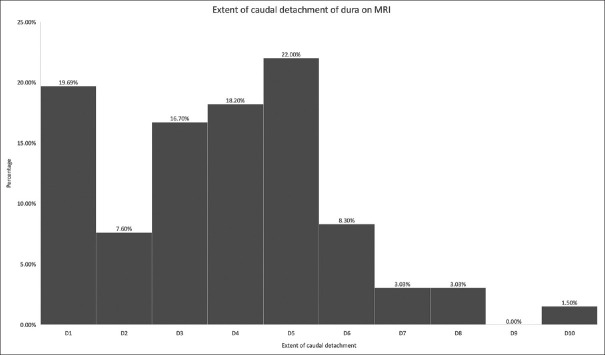
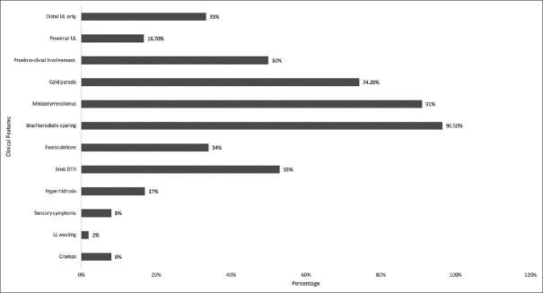
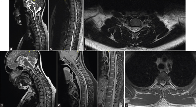
Results: One hundred and thirty-two (62.8%) patients were identified to have DD beyond the cervical spine in a cohort of 210 HD patients. The mean age at onset and duration were 18.09 ± 2.3 years (13-26) and 38.63 ± 39.9 months, respectively. Proximo-distal involvement was noted in 50% of patients, while 33% and 17% of patients had isolated distal and proximal involvement, respectively. Wasted legs were observed in three patients. Cord atrophy was present in 96.9% of patients, extending from C5 to C7. Epidural detachment and engorgement of posterior epidural venous plexus were evident in all. DD extended from C2 to D10 vertebral level. The cranial extent of DD was from C2 to C4 in 87% of cases, and the caudal extent was D1-D5 in 84% of cases, extending up to D10 in two cases.
Conclusions: The HD spectrum continues to evolve phenotypically and radiologically. The pathophysiological mechanisms and DD extend beyond the cervical spine in a large proportion of patients. This makes it important to cover a longer part of the spine during imaging. This may have implications on the management of patients, particularly those with isolated lower limb involvement.
期刊介绍:
The journal has a clinical foundation and has been utilized most by clinical neurologists for improving the practice of neurology. While the focus is on neurology in India, the journal publishes manuscripts of high value from all parts of the world. Journal publishes reviews of various types, original articles, short communications, interesting images and case reports. The journal respects the scientific submission of its authors and believes in following an expeditious double-blind peer review process and endeavors to complete the review process within scheduled time frame. A significant effort from the author and the journal perhaps enables to strike an equilibrium to meet the professional expectations of the peers in the world of scientific publication. AIAN believes in safeguarding the privacy rights of human subjects. In order to comply with it, the journal instructs all authors when uploading the manuscript to also add the ethical clearance (human/animals)/ informed consent of subject in the manuscript. This applies to the study/case report that involves animal/human subjects/human specimens e.g. extracted tooth part/soft tissue for biopsy/in vitro analysis.
 求助内容:
求助内容: 应助结果提醒方式:


