Miao Qinghao, Zhou Sheng, Yang Jun, Wang Xiaochun, Zhang Min
{"title":"基于深度学习的超声生物显微前段图像关键点定位与参数测量。","authors":"Miao Qinghao, Zhou Sheng, Yang Jun, Wang Xiaochun, Zhang Min","doi":"10.1186/s12938-025-01388-3","DOIUrl":null,"url":null,"abstract":"<p><strong>Background: </strong>Accurate measurement of anterior segment parameters is crucial for diagnosing and managing ophthalmic conditions, such as glaucoma, cataracts, and refractive errors. However, traditional clinical measurement methods are often time-consuming, labor-intensive, and susceptible to inaccuracies. With the growing potential of artificial intelligence in ophthalmic diagnostics, this study aims to develop and evaluate a deep learning model capable of automatically extracting key points and precisely measuring multiple clinically significant anterior segment parameters from ultrasound biomicroscopy (UBM) images. These parameters include central corneal thickness (CCT), anterior chamber depth (ACD), pupil diameter (PD), angle-to-angle distance (ATA), sulcus-to-sulcus distance (STS), lens thickness (LT), and crystalline lens rise (CLR).</p><p><strong>Methods: </strong>A data set of 716 UBM anterior segment images was collected from Tianjin Medical University Eye Hospital. YOLOv8 was utilized to segment four key anatomical structures: cornea-sclera, anterior chamber, pupil, and iris-ciliary body-thereby enhancing the accuracy of keypoint localization. Only images with intact posterior capsule lentis were selected to create an effective data set for parameter measurement. Ten keypoints were localized across the data set, allowing the calculation of seven essential parameters. Control experiments were conducted to evaluate the impact of segmentation on measurement accuracy, with model predictions compared against clinical gold standards.</p><p><strong>Results: </strong>The segmentation model achieved a mean IoU of 0.8836 and mPA of 0.9795. Following segmentation, the binary classification model attained an mAP of 0.9719, with a precision of 0.9260 and a recall of 0.9615. Keypoint localization exhibited a Euclidean distance error of 58.73 ± 63.04 μm, improving from the pre-segmentation error of 71.57 ± 67.36 μm. Localization mAP was 0.9826, with a precision of 0.9699, a recall of 0.9642 and an FPS of 32.64. In addition, parameter error analysis and Bland-Altman plots demonstrated improved agreement with clinical gold standards after segmentation.</p><p><strong>Conclusions: </strong>This deep learning approach for UBM image segmentation, keypoint localization, and parameter measurement is feasible, enhancing clinical diagnostic efficiency for anterior segment parameters.</p>","PeriodicalId":8927,"journal":{"name":"BioMedical Engineering OnLine","volume":"24 1","pages":"53"},"PeriodicalIF":2.9000,"publicationDate":"2025-05-06","publicationTypes":"Journal Article","fieldsOfStudy":null,"isOpenAccess":false,"openAccessPdf":"https://www.ncbi.nlm.nih.gov/pmc/articles/PMC12056989/pdf/","citationCount":"0","resultStr":"{\"title\":\"Keypoint localization and parameter measurement in ultrasound biomicroscopy anterior segment images based on deep learning.\",\"authors\":\"Miao Qinghao, Zhou Sheng, Yang Jun, Wang Xiaochun, Zhang Min\",\"doi\":\"10.1186/s12938-025-01388-3\",\"DOIUrl\":null,\"url\":null,\"abstract\":\"<p><strong>Background: </strong>Accurate measurement of anterior segment parameters is crucial for diagnosing and managing ophthalmic conditions, such as glaucoma, cataracts, and refractive errors. However, traditional clinical measurement methods are often time-consuming, labor-intensive, and susceptible to inaccuracies. With the growing potential of artificial intelligence in ophthalmic diagnostics, this study aims to develop and evaluate a deep learning model capable of automatically extracting key points and precisely measuring multiple clinically significant anterior segment parameters from ultrasound biomicroscopy (UBM) images. These parameters include central corneal thickness (CCT), anterior chamber depth (ACD), pupil diameter (PD), angle-to-angle distance (ATA), sulcus-to-sulcus distance (STS), lens thickness (LT), and crystalline lens rise (CLR).</p><p><strong>Methods: </strong>A data set of 716 UBM anterior segment images was collected from Tianjin Medical University Eye Hospital. YOLOv8 was utilized to segment four key anatomical structures: cornea-sclera, anterior chamber, pupil, and iris-ciliary body-thereby enhancing the accuracy of keypoint localization. Only images with intact posterior capsule lentis were selected to create an effective data set for parameter measurement. Ten keypoints were localized across the data set, allowing the calculation of seven essential parameters. Control experiments were conducted to evaluate the impact of segmentation on measurement accuracy, with model predictions compared against clinical gold standards.</p><p><strong>Results: </strong>The segmentation model achieved a mean IoU of 0.8836 and mPA of 0.9795. Following segmentation, the binary classification model attained an mAP of 0.9719, with a precision of 0.9260 and a recall of 0.9615. Keypoint localization exhibited a Euclidean distance error of 58.73 ± 63.04 μm, improving from the pre-segmentation error of 71.57 ± 67.36 μm. Localization mAP was 0.9826, with a precision of 0.9699, a recall of 0.9642 and an FPS of 32.64. In addition, parameter error analysis and Bland-Altman plots demonstrated improved agreement with clinical gold standards after segmentation.</p><p><strong>Conclusions: </strong>This deep learning approach for UBM image segmentation, keypoint localization, and parameter measurement is feasible, enhancing clinical diagnostic efficiency for anterior segment parameters.</p>\",\"PeriodicalId\":8927,\"journal\":{\"name\":\"BioMedical Engineering OnLine\",\"volume\":\"24 1\",\"pages\":\"53\"},\"PeriodicalIF\":2.9000,\"publicationDate\":\"2025-05-06\",\"publicationTypes\":\"Journal Article\",\"fieldsOfStudy\":null,\"isOpenAccess\":false,\"openAccessPdf\":\"https://www.ncbi.nlm.nih.gov/pmc/articles/PMC12056989/pdf/\",\"citationCount\":\"0\",\"resultStr\":null,\"platform\":\"Semanticscholar\",\"paperid\":null,\"PeriodicalName\":\"BioMedical Engineering OnLine\",\"FirstCategoryId\":\"5\",\"ListUrlMain\":\"https://doi.org/10.1186/s12938-025-01388-3\",\"RegionNum\":4,\"RegionCategory\":\"医学\",\"ArticlePicture\":[],\"TitleCN\":null,\"AbstractTextCN\":null,\"PMCID\":null,\"EPubDate\":\"\",\"PubModel\":\"\",\"JCR\":\"Q3\",\"JCRName\":\"ENGINEERING, BIOMEDICAL\",\"Score\":null,\"Total\":0}","platform":"Semanticscholar","paperid":null,"PeriodicalName":"BioMedical Engineering OnLine","FirstCategoryId":"5","ListUrlMain":"https://doi.org/10.1186/s12938-025-01388-3","RegionNum":4,"RegionCategory":"医学","ArticlePicture":[],"TitleCN":null,"AbstractTextCN":null,"PMCID":null,"EPubDate":"","PubModel":"","JCR":"Q3","JCRName":"ENGINEERING, BIOMEDICAL","Score":null,"Total":0}
Keypoint localization and parameter measurement in ultrasound biomicroscopy anterior segment images based on deep learning.
Background: Accurate measurement of anterior segment parameters is crucial for diagnosing and managing ophthalmic conditions, such as glaucoma, cataracts, and refractive errors. However, traditional clinical measurement methods are often time-consuming, labor-intensive, and susceptible to inaccuracies. With the growing potential of artificial intelligence in ophthalmic diagnostics, this study aims to develop and evaluate a deep learning model capable of automatically extracting key points and precisely measuring multiple clinically significant anterior segment parameters from ultrasound biomicroscopy (UBM) images. These parameters include central corneal thickness (CCT), anterior chamber depth (ACD), pupil diameter (PD), angle-to-angle distance (ATA), sulcus-to-sulcus distance (STS), lens thickness (LT), and crystalline lens rise (CLR).
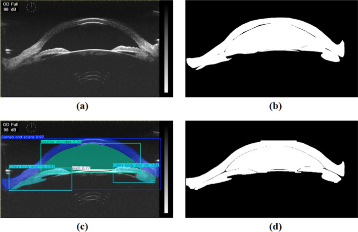
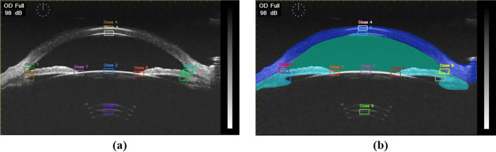
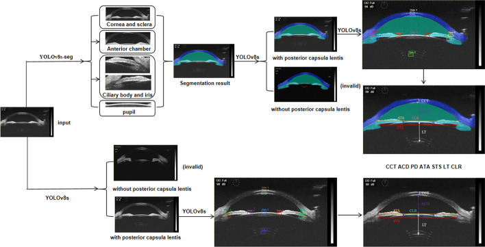
Methods: A data set of 716 UBM anterior segment images was collected from Tianjin Medical University Eye Hospital. YOLOv8 was utilized to segment four key anatomical structures: cornea-sclera, anterior chamber, pupil, and iris-ciliary body-thereby enhancing the accuracy of keypoint localization. Only images with intact posterior capsule lentis were selected to create an effective data set for parameter measurement. Ten keypoints were localized across the data set, allowing the calculation of seven essential parameters. Control experiments were conducted to evaluate the impact of segmentation on measurement accuracy, with model predictions compared against clinical gold standards.
Results: The segmentation model achieved a mean IoU of 0.8836 and mPA of 0.9795. Following segmentation, the binary classification model attained an mAP of 0.9719, with a precision of 0.9260 and a recall of 0.9615. Keypoint localization exhibited a Euclidean distance error of 58.73 ± 63.04 μm, improving from the pre-segmentation error of 71.57 ± 67.36 μm. Localization mAP was 0.9826, with a precision of 0.9699, a recall of 0.9642 and an FPS of 32.64. In addition, parameter error analysis and Bland-Altman plots demonstrated improved agreement with clinical gold standards after segmentation.
Conclusions: This deep learning approach for UBM image segmentation, keypoint localization, and parameter measurement is feasible, enhancing clinical diagnostic efficiency for anterior segment parameters.
期刊介绍:
BioMedical Engineering OnLine is an open access, peer-reviewed journal that is dedicated to publishing research in all areas of biomedical engineering.
BioMedical Engineering OnLine is aimed at readers and authors throughout the world, with an interest in using tools of the physical and data sciences and techniques in engineering to understand and solve problems in the biological and medical sciences. Topical areas include, but are not limited to:
Bioinformatics-
Bioinstrumentation-
Biomechanics-
Biomedical Devices & Instrumentation-
Biomedical Signal Processing-
Healthcare Information Systems-
Human Dynamics-
Neural Engineering-
Rehabilitation Engineering-
Biomaterials-
Biomedical Imaging & Image Processing-
BioMEMS and On-Chip Devices-
Bio-Micro/Nano Technologies-
Biomolecular Engineering-
Biosensors-
Cardiovascular Systems Engineering-
Cellular Engineering-
Clinical Engineering-
Computational Biology-
Drug Delivery Technologies-
Modeling Methodologies-
Nanomaterials and Nanotechnology in Biomedicine-
Respiratory Systems Engineering-
Robotics in Medicine-
Systems and Synthetic Biology-
Systems Biology-
Telemedicine/Smartphone Applications in Medicine-
Therapeutic Systems, Devices and Technologies-
Tissue Engineering
 求助内容:
求助内容: 应助结果提醒方式:


