Guangyuan Li, Yonghuai Wang, Bo Pang, Jun Yang, Chunyan Ma
{"title":"孤立性左束支传导阻滞患者心电图特征与亚临床左室收缩功能障碍的关系。","authors":"Guangyuan Li, Yonghuai Wang, Bo Pang, Jun Yang, Chunyan Ma","doi":"10.1186/s12947-025-00342-6","DOIUrl":null,"url":null,"abstract":"<p><strong>Background: </strong>Early identification of subclinical left ventricular (LV) systolic dysfunction (LVSD) in patients with isolated left bundle branch block (LBBB) and preserved LV ejection fraction (LVEF), termed LBBB<sub>pEF</sub>, is clinically important. Electrocardiography (ECG) has been proposed as a potential screening tool for detecting subclinical LVSD in LBBB<sub>pEF</sub> patients, but its effectiveness has not been fully validated. This study investigated the relationships between specific ECG characteristics and subclinical LVSD in LBBB<sub>pEF</sub> patients.</p><p><strong>Methods: </strong>The study included 111 patients with LBBB<sub>pEF</sub>. Two-dimensional speckle-tracking echocardiography was used to derive the LV global longitudinal strain (LV GLS), with LV GLS>-20% indicating subclinical LVSD. The recorded ECG characteristics included heart rate, QRS duration, P-R duration, QRS morphology, T-wave morphology, the presence of QS patterns, and discordant LBBB, among others. The presence of QS patterns was defined as the absence of R-waves in lead V1 (or R-waves < 1 mm with a scale of 10 mm/mV). Discordant LBBB was defined as an inconsistency between the T wave and QRS complex in leads I, V5, and V6.</p><p><strong>Results: </strong>Among the patients, 52 exhibited subclinical LVSD. Compared with those with normal LV systolic function, patients with subclinical LVSD had longer QRS durations, a higher frequency of QS patterns, and more instances of discordant LBBB. A QRS duration of 153 ms was identified as the optimal cut-off for detecting subclinical LVSD, with a sensitivity of 75.00% and specificity of 72.88%. The combination of QRS duration, the presence of QS patterns, and discordant LBBB produced the highest area under the curve of 0.82. Incorporating the presence of QS patterns and discordant LBBB into the QRS duration model increased the integrated discriminant index from 0.07 to 0.15.</p><p><strong>Conclusions: </strong>QRS duration, the presence of QS patterns, and discordant LBBB are independent predictors of subclinical LVSD in patients with LBBB<sub>pEF</sub>. An integrated ECG assessment may offer a straightforward screening method for identifying subclinical LVSD in this population.</p>","PeriodicalId":9613,"journal":{"name":"Cardiovascular Ultrasound","volume":"23 1","pages":"7"},"PeriodicalIF":1.6000,"publicationDate":"2025-05-01","publicationTypes":"Journal Article","fieldsOfStudy":null,"isOpenAccess":false,"openAccessPdf":"https://www.ncbi.nlm.nih.gov/pmc/articles/PMC12044813/pdf/","citationCount":"0","resultStr":"{\"title\":\"Relationship between electrocardiographic characteristics and subclinical left ventricular systolic dysfunction in isolated left bundle branch block patients.\",\"authors\":\"Guangyuan Li, Yonghuai Wang, Bo Pang, Jun Yang, Chunyan Ma\",\"doi\":\"10.1186/s12947-025-00342-6\",\"DOIUrl\":null,\"url\":null,\"abstract\":\"<p><strong>Background: </strong>Early identification of subclinical left ventricular (LV) systolic dysfunction (LVSD) in patients with isolated left bundle branch block (LBBB) and preserved LV ejection fraction (LVEF), termed LBBB<sub>pEF</sub>, is clinically important. Electrocardiography (ECG) has been proposed as a potential screening tool for detecting subclinical LVSD in LBBB<sub>pEF</sub> patients, but its effectiveness has not been fully validated. This study investigated the relationships between specific ECG characteristics and subclinical LVSD in LBBB<sub>pEF</sub> patients.</p><p><strong>Methods: </strong>The study included 111 patients with LBBB<sub>pEF</sub>. Two-dimensional speckle-tracking echocardiography was used to derive the LV global longitudinal strain (LV GLS), with LV GLS>-20% indicating subclinical LVSD. The recorded ECG characteristics included heart rate, QRS duration, P-R duration, QRS morphology, T-wave morphology, the presence of QS patterns, and discordant LBBB, among others. The presence of QS patterns was defined as the absence of R-waves in lead V1 (or R-waves < 1 mm with a scale of 10 mm/mV). Discordant LBBB was defined as an inconsistency between the T wave and QRS complex in leads I, V5, and V6.</p><p><strong>Results: </strong>Among the patients, 52 exhibited subclinical LVSD. Compared with those with normal LV systolic function, patients with subclinical LVSD had longer QRS durations, a higher frequency of QS patterns, and more instances of discordant LBBB. A QRS duration of 153 ms was identified as the optimal cut-off for detecting subclinical LVSD, with a sensitivity of 75.00% and specificity of 72.88%. The combination of QRS duration, the presence of QS patterns, and discordant LBBB produced the highest area under the curve of 0.82. Incorporating the presence of QS patterns and discordant LBBB into the QRS duration model increased the integrated discriminant index from 0.07 to 0.15.</p><p><strong>Conclusions: </strong>QRS duration, the presence of QS patterns, and discordant LBBB are independent predictors of subclinical LVSD in patients with LBBB<sub>pEF</sub>. An integrated ECG assessment may offer a straightforward screening method for identifying subclinical LVSD in this population.</p>\",\"PeriodicalId\":9613,\"journal\":{\"name\":\"Cardiovascular Ultrasound\",\"volume\":\"23 1\",\"pages\":\"7\"},\"PeriodicalIF\":1.6000,\"publicationDate\":\"2025-05-01\",\"publicationTypes\":\"Journal Article\",\"fieldsOfStudy\":null,\"isOpenAccess\":false,\"openAccessPdf\":\"https://www.ncbi.nlm.nih.gov/pmc/articles/PMC12044813/pdf/\",\"citationCount\":\"0\",\"resultStr\":null,\"platform\":\"Semanticscholar\",\"paperid\":null,\"PeriodicalName\":\"Cardiovascular Ultrasound\",\"FirstCategoryId\":\"3\",\"ListUrlMain\":\"https://doi.org/10.1186/s12947-025-00342-6\",\"RegionNum\":3,\"RegionCategory\":\"医学\",\"ArticlePicture\":[],\"TitleCN\":null,\"AbstractTextCN\":null,\"PMCID\":null,\"EPubDate\":\"\",\"PubModel\":\"\",\"JCR\":\"Q3\",\"JCRName\":\"CARDIAC & CARDIOVASCULAR SYSTEMS\",\"Score\":null,\"Total\":0}","platform":"Semanticscholar","paperid":null,"PeriodicalName":"Cardiovascular Ultrasound","FirstCategoryId":"3","ListUrlMain":"https://doi.org/10.1186/s12947-025-00342-6","RegionNum":3,"RegionCategory":"医学","ArticlePicture":[],"TitleCN":null,"AbstractTextCN":null,"PMCID":null,"EPubDate":"","PubModel":"","JCR":"Q3","JCRName":"CARDIAC & CARDIOVASCULAR SYSTEMS","Score":null,"Total":0}
Relationship between electrocardiographic characteristics and subclinical left ventricular systolic dysfunction in isolated left bundle branch block patients.
Background: Early identification of subclinical left ventricular (LV) systolic dysfunction (LVSD) in patients with isolated left bundle branch block (LBBB) and preserved LV ejection fraction (LVEF), termed LBBBpEF, is clinically important. Electrocardiography (ECG) has been proposed as a potential screening tool for detecting subclinical LVSD in LBBBpEF patients, but its effectiveness has not been fully validated. This study investigated the relationships between specific ECG characteristics and subclinical LVSD in LBBBpEF patients.
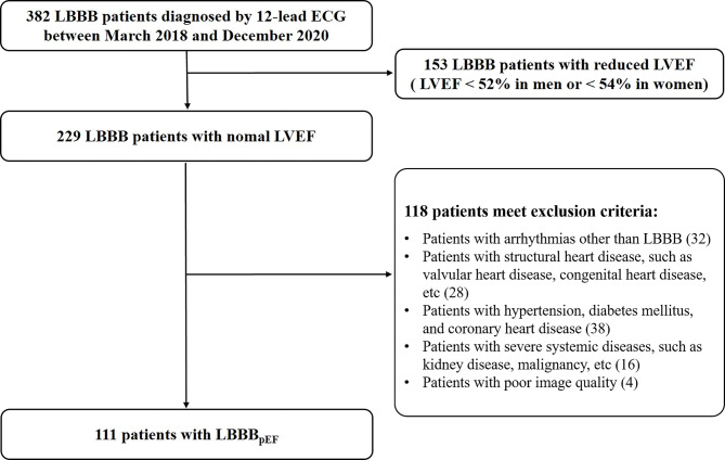
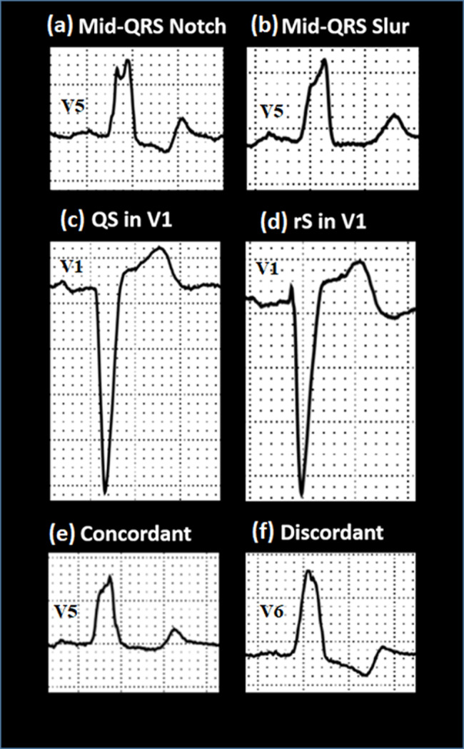
Methods: The study included 111 patients with LBBBpEF. Two-dimensional speckle-tracking echocardiography was used to derive the LV global longitudinal strain (LV GLS), with LV GLS>-20% indicating subclinical LVSD. The recorded ECG characteristics included heart rate, QRS duration, P-R duration, QRS morphology, T-wave morphology, the presence of QS patterns, and discordant LBBB, among others. The presence of QS patterns was defined as the absence of R-waves in lead V1 (or R-waves < 1 mm with a scale of 10 mm/mV). Discordant LBBB was defined as an inconsistency between the T wave and QRS complex in leads I, V5, and V6.
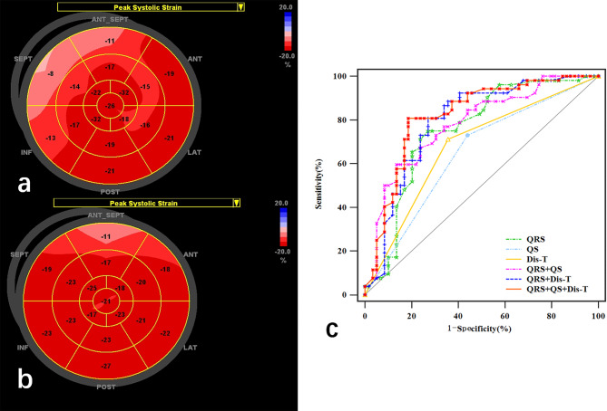
Results: Among the patients, 52 exhibited subclinical LVSD. Compared with those with normal LV systolic function, patients with subclinical LVSD had longer QRS durations, a higher frequency of QS patterns, and more instances of discordant LBBB. A QRS duration of 153 ms was identified as the optimal cut-off for detecting subclinical LVSD, with a sensitivity of 75.00% and specificity of 72.88%. The combination of QRS duration, the presence of QS patterns, and discordant LBBB produced the highest area under the curve of 0.82. Incorporating the presence of QS patterns and discordant LBBB into the QRS duration model increased the integrated discriminant index from 0.07 to 0.15.
Conclusions: QRS duration, the presence of QS patterns, and discordant LBBB are independent predictors of subclinical LVSD in patients with LBBBpEF. An integrated ECG assessment may offer a straightforward screening method for identifying subclinical LVSD in this population.
期刊介绍:
Cardiovascular Ultrasound is an online journal, publishing peer-reviewed: original research; authoritative reviews; case reports on challenging and/or unusual diagnostic aspects; and expert opinions on new techniques and technologies. We are particularly interested in articles that include relevant images or video files, which provide an additional dimension to published articles and enhance understanding.
As an open access journal, Cardiovascular Ultrasound ensures high visibility for authors in addition to providing an up-to-date and freely available resource for the community. The journal welcomes discussion, and provides a forum for publishing opinion and debate ranging from biology to engineering to clinical echocardiography, with both speed and versatility.
 求助内容:
求助内容: 应助结果提醒方式:


