Mohamed M Khodeiry, Mohammad Ayoubi, Christopher A Dorizas, Carlos E Mendoza-Santiesteban, Maja Kostic
{"title":"玻璃体乳头牵引引起视神经头抬高。","authors":"Mohamed M Khodeiry, Mohammad Ayoubi, Christopher A Dorizas, Carlos E Mendoza-Santiesteban, Maja Kostic","doi":"10.1155/crop/3136288","DOIUrl":null,"url":null,"abstract":"<p><p><b>Purpose:</b> The purpose of the study is to describe a case of vitreopapillary traction causing optic nerve head elevation. <b>Observations:</b> This case report describes a 64-year-old male who presented with left cloudy vision for 3 days. Dilated fundus exam showed normal right optic nerve with glial tissue nasally and left optic nerve head elevation and peripapillary hemorrhages in the left eye. Magnetic resonance imaging of the brain and orbits, erythrocyte sedimentation rate, and C-reactive protein were normal. Optical coherence tomography showed bilateral dense vitreous adhesions to the optic disc nasally causing traction and optic nerve head elevation of the left eye. The patient was diagnosed with vitreopapillary traction causing optic nerve head elevation, and observation was recommended. <b>Conclusions and Importance:</b> This case highlights the importance of clinical examination and ancillary testing in differentiating etiologies of optic disc elevation.</p>","PeriodicalId":9603,"journal":{"name":"Case Reports in Ophthalmological Medicine","volume":"2025 ","pages":"3136288"},"PeriodicalIF":0.4000,"publicationDate":"2025-04-23","publicationTypes":"Journal Article","fieldsOfStudy":null,"isOpenAccess":false,"openAccessPdf":"https://www.ncbi.nlm.nih.gov/pmc/articles/PMC12043439/pdf/","citationCount":"0","resultStr":"{\"title\":\"Vitreopapillary Traction Causing Optic Nerve Head Elevation.\",\"authors\":\"Mohamed M Khodeiry, Mohammad Ayoubi, Christopher A Dorizas, Carlos E Mendoza-Santiesteban, Maja Kostic\",\"doi\":\"10.1155/crop/3136288\",\"DOIUrl\":null,\"url\":null,\"abstract\":\"<p><p><b>Purpose:</b> The purpose of the study is to describe a case of vitreopapillary traction causing optic nerve head elevation. <b>Observations:</b> This case report describes a 64-year-old male who presented with left cloudy vision for 3 days. Dilated fundus exam showed normal right optic nerve with glial tissue nasally and left optic nerve head elevation and peripapillary hemorrhages in the left eye. Magnetic resonance imaging of the brain and orbits, erythrocyte sedimentation rate, and C-reactive protein were normal. Optical coherence tomography showed bilateral dense vitreous adhesions to the optic disc nasally causing traction and optic nerve head elevation of the left eye. The patient was diagnosed with vitreopapillary traction causing optic nerve head elevation, and observation was recommended. <b>Conclusions and Importance:</b> This case highlights the importance of clinical examination and ancillary testing in differentiating etiologies of optic disc elevation.</p>\",\"PeriodicalId\":9603,\"journal\":{\"name\":\"Case Reports in Ophthalmological Medicine\",\"volume\":\"2025 \",\"pages\":\"3136288\"},\"PeriodicalIF\":0.4000,\"publicationDate\":\"2025-04-23\",\"publicationTypes\":\"Journal Article\",\"fieldsOfStudy\":null,\"isOpenAccess\":false,\"openAccessPdf\":\"https://www.ncbi.nlm.nih.gov/pmc/articles/PMC12043439/pdf/\",\"citationCount\":\"0\",\"resultStr\":null,\"platform\":\"Semanticscholar\",\"paperid\":null,\"PeriodicalName\":\"Case Reports in Ophthalmological Medicine\",\"FirstCategoryId\":\"1085\",\"ListUrlMain\":\"https://doi.org/10.1155/crop/3136288\",\"RegionNum\":0,\"RegionCategory\":null,\"ArticlePicture\":[],\"TitleCN\":null,\"AbstractTextCN\":null,\"PMCID\":null,\"EPubDate\":\"2025/1/1 0:00:00\",\"PubModel\":\"eCollection\",\"JCR\":\"Q4\",\"JCRName\":\"OPHTHALMOLOGY\",\"Score\":null,\"Total\":0}","platform":"Semanticscholar","paperid":null,"PeriodicalName":"Case Reports in Ophthalmological Medicine","FirstCategoryId":"1085","ListUrlMain":"https://doi.org/10.1155/crop/3136288","RegionNum":0,"RegionCategory":null,"ArticlePicture":[],"TitleCN":null,"AbstractTextCN":null,"PMCID":null,"EPubDate":"2025/1/1 0:00:00","PubModel":"eCollection","JCR":"Q4","JCRName":"OPHTHALMOLOGY","Score":null,"Total":0}
Vitreopapillary Traction Causing Optic Nerve Head Elevation.
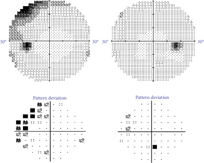
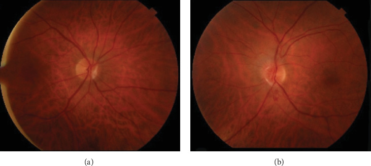
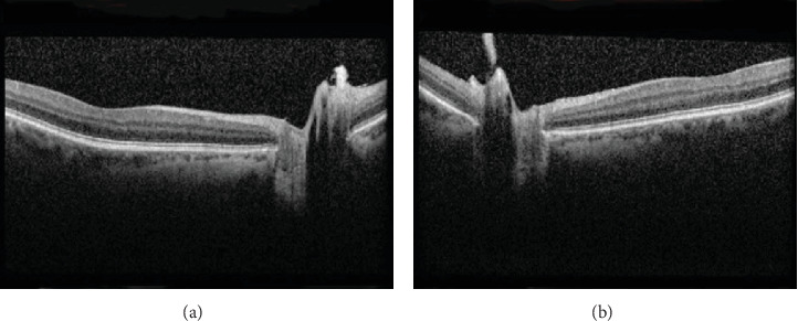
Purpose: The purpose of the study is to describe a case of vitreopapillary traction causing optic nerve head elevation. Observations: This case report describes a 64-year-old male who presented with left cloudy vision for 3 days. Dilated fundus exam showed normal right optic nerve with glial tissue nasally and left optic nerve head elevation and peripapillary hemorrhages in the left eye. Magnetic resonance imaging of the brain and orbits, erythrocyte sedimentation rate, and C-reactive protein were normal. Optical coherence tomography showed bilateral dense vitreous adhesions to the optic disc nasally causing traction and optic nerve head elevation of the left eye. The patient was diagnosed with vitreopapillary traction causing optic nerve head elevation, and observation was recommended. Conclusions and Importance: This case highlights the importance of clinical examination and ancillary testing in differentiating etiologies of optic disc elevation.

 求助内容:
求助内容: 应助结果提醒方式:


