Bruna Nichele da Rosa, Paula Andryelly Gomes Giendruczak, Marina Ziegler Frantz, Matias Noll, Cláudia Tarragô Candotti
{"title":"三种摄影测量方法在矢状面评估膝关节直线的同时有效性。","authors":"Bruna Nichele da Rosa, Paula Andryelly Gomes Giendruczak, Marina Ziegler Frantz, Matias Noll, Cláudia Tarragô Candotti","doi":"10.3390/mps8020041","DOIUrl":null,"url":null,"abstract":"<p><p><i>Background</i>: Evidence supporting the validity of photogrammetry for assessing body segment alignment remains limited, with most studies focusing on spinal evaluation. Thus, there is a lack of robust research examining its use for other body segments such as the lower limbs. <i>Objective</i>: This study aimed to evaluate the concurrent validity of three photogrammetric methods for measuring knee alignment in the sagittal plane with and without corrections for potential rotational deviations in the participant's thigh and leg. <i>Methods</i>: A total of 21 adults underwent sequential evaluations involving panoramic radiography of the lower limbs and photogrammetry at a private radiology clinic. Photogrammetric analysis involved identifying the following anatomical landmarks: the greater trochanter of the femur (GTF), the lateral condyle of the femur (LCF), the head of the fibula (HF), and lateral malleolus (LM). Three photogrammetric methods were employed: (1) the condylar angle (CA) defined by the GTF, LCF, and LM points; (2) the fibula head angle (FHA) defined by the GTF, HF, and LM points; and (3) the four-point angle (4PA) incorporating the GTF, LCF, HF, and LM. Concurrent validity was assessed using correlation analysis, agreement with radiographic measurements, and the root mean square error (RMSE). Each photogrammetric method was tested using raw (CA, FHA, and 4PA) and corrected (CAcorr, FHAcorr, and 4PAcorr) values, accounting for thigh and/or leg rotational deviations. <i>Results</i>: Correcting for thigh and leg rotations significantly improved the validity metrics for all methods. The best performance was observed with the corrected condylar angle (CAcorr: r = 0.746; adjusted r<sup>2</sup> = 0.533; RMSE = 2.9°) and the corrected four-point angle (4PAcorr: r = 0.733; adjusted r<sup>2</sup> = 0.513; RMSE = 3.0°); however, the measurements presented proportional errors, possible due the method of assessment of rotations. <i>Conclusions</i>: The findings validate the evaluated photogrammetric methods for assessing sagittal knee alignment. Accounting for thigh and leg rotational deviations is critical for achieving accurate measurements, raising the need of accurate tools for measuring rotational changes in the lower limbs to avoid errors.</p>","PeriodicalId":18715,"journal":{"name":"Methods and Protocols","volume":"8 2","pages":""},"PeriodicalIF":2.0000,"publicationDate":"2025-04-14","publicationTypes":"Journal Article","fieldsOfStudy":null,"isOpenAccess":false,"openAccessPdf":"https://www.ncbi.nlm.nih.gov/pmc/articles/PMC12029935/pdf/","citationCount":"0","resultStr":"{\"title\":\"Concurrent Validity of Three Photogrammetric Methods for Assessing Knee Alignment in Sagittal Plane.\",\"authors\":\"Bruna Nichele da Rosa, Paula Andryelly Gomes Giendruczak, Marina Ziegler Frantz, Matias Noll, Cláudia Tarragô Candotti\",\"doi\":\"10.3390/mps8020041\",\"DOIUrl\":null,\"url\":null,\"abstract\":\"<p><p><i>Background</i>: Evidence supporting the validity of photogrammetry for assessing body segment alignment remains limited, with most studies focusing on spinal evaluation. Thus, there is a lack of robust research examining its use for other body segments such as the lower limbs. <i>Objective</i>: This study aimed to evaluate the concurrent validity of three photogrammetric methods for measuring knee alignment in the sagittal plane with and without corrections for potential rotational deviations in the participant's thigh and leg. <i>Methods</i>: A total of 21 adults underwent sequential evaluations involving panoramic radiography of the lower limbs and photogrammetry at a private radiology clinic. Photogrammetric analysis involved identifying the following anatomical landmarks: the greater trochanter of the femur (GTF), the lateral condyle of the femur (LCF), the head of the fibula (HF), and lateral malleolus (LM). Three photogrammetric methods were employed: (1) the condylar angle (CA) defined by the GTF, LCF, and LM points; (2) the fibula head angle (FHA) defined by the GTF, HF, and LM points; and (3) the four-point angle (4PA) incorporating the GTF, LCF, HF, and LM. Concurrent validity was assessed using correlation analysis, agreement with radiographic measurements, and the root mean square error (RMSE). Each photogrammetric method was tested using raw (CA, FHA, and 4PA) and corrected (CAcorr, FHAcorr, and 4PAcorr) values, accounting for thigh and/or leg rotational deviations. <i>Results</i>: Correcting for thigh and leg rotations significantly improved the validity metrics for all methods. The best performance was observed with the corrected condylar angle (CAcorr: r = 0.746; adjusted r<sup>2</sup> = 0.533; RMSE = 2.9°) and the corrected four-point angle (4PAcorr: r = 0.733; adjusted r<sup>2</sup> = 0.513; RMSE = 3.0°); however, the measurements presented proportional errors, possible due the method of assessment of rotations. <i>Conclusions</i>: The findings validate the evaluated photogrammetric methods for assessing sagittal knee alignment. Accounting for thigh and leg rotational deviations is critical for achieving accurate measurements, raising the need of accurate tools for measuring rotational changes in the lower limbs to avoid errors.</p>\",\"PeriodicalId\":18715,\"journal\":{\"name\":\"Methods and Protocols\",\"volume\":\"8 2\",\"pages\":\"\"},\"PeriodicalIF\":2.0000,\"publicationDate\":\"2025-04-14\",\"publicationTypes\":\"Journal Article\",\"fieldsOfStudy\":null,\"isOpenAccess\":false,\"openAccessPdf\":\"https://www.ncbi.nlm.nih.gov/pmc/articles/PMC12029935/pdf/\",\"citationCount\":\"0\",\"resultStr\":null,\"platform\":\"Semanticscholar\",\"paperid\":null,\"PeriodicalName\":\"Methods and Protocols\",\"FirstCategoryId\":\"1085\",\"ListUrlMain\":\"https://doi.org/10.3390/mps8020041\",\"RegionNum\":0,\"RegionCategory\":null,\"ArticlePicture\":[],\"TitleCN\":null,\"AbstractTextCN\":null,\"PMCID\":null,\"EPubDate\":\"\",\"PubModel\":\"\",\"JCR\":\"Q3\",\"JCRName\":\"BIOCHEMICAL RESEARCH METHODS\",\"Score\":null,\"Total\":0}","platform":"Semanticscholar","paperid":null,"PeriodicalName":"Methods and Protocols","FirstCategoryId":"1085","ListUrlMain":"https://doi.org/10.3390/mps8020041","RegionNum":0,"RegionCategory":null,"ArticlePicture":[],"TitleCN":null,"AbstractTextCN":null,"PMCID":null,"EPubDate":"","PubModel":"","JCR":"Q3","JCRName":"BIOCHEMICAL RESEARCH METHODS","Score":null,"Total":0}
引用次数: 0
摘要
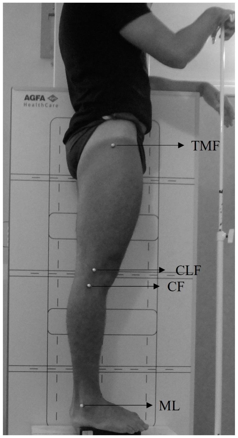
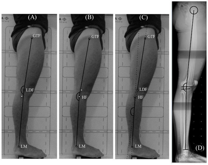
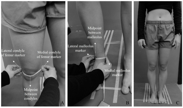
背景:支持摄影测量评估身体节段对齐有效性的证据仍然有限,大多数研究集中在脊柱评估上。因此,缺乏强有力的研究来检验它在其他身体部位(如下肢)的应用。目的:本研究旨在评估三种测量膝关节矢状面对齐的摄影测量方法的同时有效性,并对参与者大腿和腿部的潜在旋转偏差进行校正。方法:共有21名成年人在一家私人放射诊所接受了包括下肢全景x线摄影和摄影测量在内的连续评估。摄影测量分析包括确定以下解剖标志:股骨大转子(GTF)、股骨外侧髁(LCF)、腓骨头(HF)和外踝(LM)。采用三种摄影测量方法:(1)由GTF、LCF和LM点定义的髁角(CA);(2)由GTF、HF、LM点定义的腓骨头角(FHA);(3)包含GTF、LCF、HF和LM的四点角(4PA)。同时效度评估采用相关分析,与x线测量的一致性和均方根误差(RMSE)。每种摄影测量方法使用原始(CA、FHA和4PA)和校正(CAcorr、FHAcorr和4PAcorr)值进行测试,考虑大腿和/或腿部旋转偏差。结果:纠正大腿和腿部旋转显著提高了所有方法的效度指标。矫正后的髁角效果最佳(CAcorr: r = 0.746;调整后r2 = 0.533;RMSE = 2.9°)和修正后的四点角(4PAcorr: r = 0.733;调整后r2 = 0.513;Rmse = 3.0°);然而,测量结果存在比例误差,可能是由于评估旋转的方法。结论:研究结果验证了评估膝关节矢状位对齐的摄影测量方法。考虑大腿和腿部旋转偏差是实现准确测量的关键,提高了对测量下肢旋转变化的精确工具的需求,以避免误差。
Concurrent Validity of Three Photogrammetric Methods for Assessing Knee Alignment in Sagittal Plane.
Background: Evidence supporting the validity of photogrammetry for assessing body segment alignment remains limited, with most studies focusing on spinal evaluation. Thus, there is a lack of robust research examining its use for other body segments such as the lower limbs. Objective: This study aimed to evaluate the concurrent validity of three photogrammetric methods for measuring knee alignment in the sagittal plane with and without corrections for potential rotational deviations in the participant's thigh and leg. Methods: A total of 21 adults underwent sequential evaluations involving panoramic radiography of the lower limbs and photogrammetry at a private radiology clinic. Photogrammetric analysis involved identifying the following anatomical landmarks: the greater trochanter of the femur (GTF), the lateral condyle of the femur (LCF), the head of the fibula (HF), and lateral malleolus (LM). Three photogrammetric methods were employed: (1) the condylar angle (CA) defined by the GTF, LCF, and LM points; (2) the fibula head angle (FHA) defined by the GTF, HF, and LM points; and (3) the four-point angle (4PA) incorporating the GTF, LCF, HF, and LM. Concurrent validity was assessed using correlation analysis, agreement with radiographic measurements, and the root mean square error (RMSE). Each photogrammetric method was tested using raw (CA, FHA, and 4PA) and corrected (CAcorr, FHAcorr, and 4PAcorr) values, accounting for thigh and/or leg rotational deviations. Results: Correcting for thigh and leg rotations significantly improved the validity metrics for all methods. The best performance was observed with the corrected condylar angle (CAcorr: r = 0.746; adjusted r2 = 0.533; RMSE = 2.9°) and the corrected four-point angle (4PAcorr: r = 0.733; adjusted r2 = 0.513; RMSE = 3.0°); however, the measurements presented proportional errors, possible due the method of assessment of rotations. Conclusions: The findings validate the evaluated photogrammetric methods for assessing sagittal knee alignment. Accounting for thigh and leg rotational deviations is critical for achieving accurate measurements, raising the need of accurate tools for measuring rotational changes in the lower limbs to avoid errors.

 求助内容:
求助内容: 应助结果提醒方式:


