Marcussi Palata Rezende, Fernanda Atoui Faria, Daniel Prado Beraldo, Julia Polido, Rubens Belfort, Thiago Cabral
{"title":"早期糖尿病黄斑水肿患者扫描源OCT和OCT血管造影生物标志物的前瞻性和二分类研究。","authors":"Marcussi Palata Rezende, Fernanda Atoui Faria, Daniel Prado Beraldo, Julia Polido, Rubens Belfort, Thiago Cabral","doi":"10.1186/s40942-025-00672-7","DOIUrl":null,"url":null,"abstract":"<p><strong>Background: </strong>We used state-of-the-art high-resolution retinal imaging to explore the treatment (loading dose of aflibercept) of diabetic macular edema (DME) among treatment-naive patients. Swept-source (SS) OCT and OCT-Angiography (SS-OCTA) were performed, and a dichotomous analysis was conducted to compare responders and treatment-resistant patients (responsive and resistant). Furthermore, treatment responses were evaluated based on the subdivision of choroidal thickness.</p><p><strong>Materials and methods: </strong>This prospective, noncomparative, interventional case series study examined the following biomarkers: best-corrected visual acuity (BCVA), central macular thickness (CMT), central choroidal thickness (CCT), avascular area of the superficial plexus (AASP), avascular area of the deep plexus (AADP), and vessel density (VD). Data from the baseline and 4-month examinations were compared.</p><p><strong>Results: </strong>Twenty-eight eyes from 25 patients were included. Significant improvements were observed in BCVA (0.7250 ± 0.23 to 0.3957 ± 0.21; p < 0.000), CMT µm (339.04 ± 66.19 to 265.21 ± 55.75; p < 0.000), CCT µm (221.71 ± 69.69 to 209.07 ± 70.92; p < 0.000), VD (17.90 ± 7.82 to 15.35 ± 5.80; p < 0.038), AASP µm<sup>2</sup> (235,374 ± 91,299 to 157,326 ± 77,815; p < 0.000) and AADP µm<sup>2</sup> (996,335 ± 1,000,047 to 362,161 ± 277,225; p < 0.000). Dichotomous analysis revealed that 15 patients were responsive (53.57%), and 13 resistant (46.43%). There were no significant differences between any of the pretreatment biomarkers. In the subdivision of choroidal thickness, which ranged from 211 to 270 µm (group 3), we found greater reductions in the CCT, AADP and CD. The choroidal thickness ranged from 181 to 210 µm (group 2): BCVA and AASP exhibited the greatest reductions.</p><p><strong>Conclusion: </strong>BCVA, CMT, CCT, AASP, AADP and VD were improved after treatment. The pretreatment biomarkers did not predict treatment response between the responsive and resistant. Regarding choroidal stratification, values within the normal range of CCT showed the greatest reductions, indicating that these values may be more responsive to treatment. Notably, this is the first study to analyze biomarkers provided by SS OCT and OCTA, stratify the choroid, and perform a dichotomous analysis.</p>","PeriodicalId":14289,"journal":{"name":"International Journal of Retina and Vitreous","volume":"11 1","pages":"51"},"PeriodicalIF":2.4000,"publicationDate":"2025-04-22","publicationTypes":"Journal Article","fieldsOfStudy":null,"isOpenAccess":false,"openAccessPdf":"https://www.ncbi.nlm.nih.gov/pmc/articles/PMC12016437/pdf/","citationCount":"0","resultStr":"{\"title\":\"Prospective and dichotomous study of biomarkers with swept-source OCT and OCT-angiography in naive patients with diabetic macular edema.\",\"authors\":\"Marcussi Palata Rezende, Fernanda Atoui Faria, Daniel Prado Beraldo, Julia Polido, Rubens Belfort, Thiago Cabral\",\"doi\":\"10.1186/s40942-025-00672-7\",\"DOIUrl\":null,\"url\":null,\"abstract\":\"<p><strong>Background: </strong>We used state-of-the-art high-resolution retinal imaging to explore the treatment (loading dose of aflibercept) of diabetic macular edema (DME) among treatment-naive patients. Swept-source (SS) OCT and OCT-Angiography (SS-OCTA) were performed, and a dichotomous analysis was conducted to compare responders and treatment-resistant patients (responsive and resistant). Furthermore, treatment responses were evaluated based on the subdivision of choroidal thickness.</p><p><strong>Materials and methods: </strong>This prospective, noncomparative, interventional case series study examined the following biomarkers: best-corrected visual acuity (BCVA), central macular thickness (CMT), central choroidal thickness (CCT), avascular area of the superficial plexus (AASP), avascular area of the deep plexus (AADP), and vessel density (VD). Data from the baseline and 4-month examinations were compared.</p><p><strong>Results: </strong>Twenty-eight eyes from 25 patients were included. Significant improvements were observed in BCVA (0.7250 ± 0.23 to 0.3957 ± 0.21; p < 0.000), CMT µm (339.04 ± 66.19 to 265.21 ± 55.75; p < 0.000), CCT µm (221.71 ± 69.69 to 209.07 ± 70.92; p < 0.000), VD (17.90 ± 7.82 to 15.35 ± 5.80; p < 0.038), AASP µm<sup>2</sup> (235,374 ± 91,299 to 157,326 ± 77,815; p < 0.000) and AADP µm<sup>2</sup> (996,335 ± 1,000,047 to 362,161 ± 277,225; p < 0.000). Dichotomous analysis revealed that 15 patients were responsive (53.57%), and 13 resistant (46.43%). There were no significant differences between any of the pretreatment biomarkers. In the subdivision of choroidal thickness, which ranged from 211 to 270 µm (group 3), we found greater reductions in the CCT, AADP and CD. The choroidal thickness ranged from 181 to 210 µm (group 2): BCVA and AASP exhibited the greatest reductions.</p><p><strong>Conclusion: </strong>BCVA, CMT, CCT, AASP, AADP and VD were improved after treatment. The pretreatment biomarkers did not predict treatment response between the responsive and resistant. Regarding choroidal stratification, values within the normal range of CCT showed the greatest reductions, indicating that these values may be more responsive to treatment. Notably, this is the first study to analyze biomarkers provided by SS OCT and OCTA, stratify the choroid, and perform a dichotomous analysis.</p>\",\"PeriodicalId\":14289,\"journal\":{\"name\":\"International Journal of Retina and Vitreous\",\"volume\":\"11 1\",\"pages\":\"51\"},\"PeriodicalIF\":2.4000,\"publicationDate\":\"2025-04-22\",\"publicationTypes\":\"Journal Article\",\"fieldsOfStudy\":null,\"isOpenAccess\":false,\"openAccessPdf\":\"https://www.ncbi.nlm.nih.gov/pmc/articles/PMC12016437/pdf/\",\"citationCount\":\"0\",\"resultStr\":null,\"platform\":\"Semanticscholar\",\"paperid\":null,\"PeriodicalName\":\"International Journal of Retina and Vitreous\",\"FirstCategoryId\":\"1085\",\"ListUrlMain\":\"https://doi.org/10.1186/s40942-025-00672-7\",\"RegionNum\":0,\"RegionCategory\":null,\"ArticlePicture\":[],\"TitleCN\":null,\"AbstractTextCN\":null,\"PMCID\":null,\"EPubDate\":\"\",\"PubModel\":\"\",\"JCR\":\"Q2\",\"JCRName\":\"OPHTHALMOLOGY\",\"Score\":null,\"Total\":0}","platform":"Semanticscholar","paperid":null,"PeriodicalName":"International Journal of Retina and Vitreous","FirstCategoryId":"1085","ListUrlMain":"https://doi.org/10.1186/s40942-025-00672-7","RegionNum":0,"RegionCategory":null,"ArticlePicture":[],"TitleCN":null,"AbstractTextCN":null,"PMCID":null,"EPubDate":"","PubModel":"","JCR":"Q2","JCRName":"OPHTHALMOLOGY","Score":null,"Total":0}
Prospective and dichotomous study of biomarkers with swept-source OCT and OCT-angiography in naive patients with diabetic macular edema.
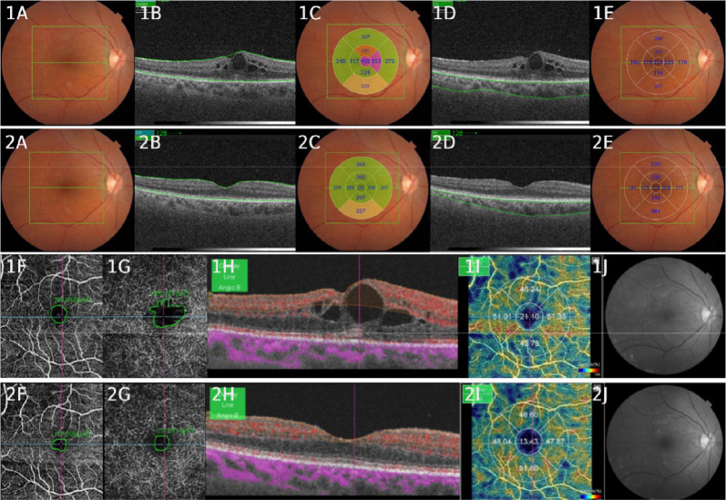
Background: We used state-of-the-art high-resolution retinal imaging to explore the treatment (loading dose of aflibercept) of diabetic macular edema (DME) among treatment-naive patients. Swept-source (SS) OCT and OCT-Angiography (SS-OCTA) were performed, and a dichotomous analysis was conducted to compare responders and treatment-resistant patients (responsive and resistant). Furthermore, treatment responses were evaluated based on the subdivision of choroidal thickness.
Materials and methods: This prospective, noncomparative, interventional case series study examined the following biomarkers: best-corrected visual acuity (BCVA), central macular thickness (CMT), central choroidal thickness (CCT), avascular area of the superficial plexus (AASP), avascular area of the deep plexus (AADP), and vessel density (VD). Data from the baseline and 4-month examinations were compared.
Results: Twenty-eight eyes from 25 patients were included. Significant improvements were observed in BCVA (0.7250 ± 0.23 to 0.3957 ± 0.21; p < 0.000), CMT µm (339.04 ± 66.19 to 265.21 ± 55.75; p < 0.000), CCT µm (221.71 ± 69.69 to 209.07 ± 70.92; p < 0.000), VD (17.90 ± 7.82 to 15.35 ± 5.80; p < 0.038), AASP µm2 (235,374 ± 91,299 to 157,326 ± 77,815; p < 0.000) and AADP µm2 (996,335 ± 1,000,047 to 362,161 ± 277,225; p < 0.000). Dichotomous analysis revealed that 15 patients were responsive (53.57%), and 13 resistant (46.43%). There were no significant differences between any of the pretreatment biomarkers. In the subdivision of choroidal thickness, which ranged from 211 to 270 µm (group 3), we found greater reductions in the CCT, AADP and CD. The choroidal thickness ranged from 181 to 210 µm (group 2): BCVA and AASP exhibited the greatest reductions.
Conclusion: BCVA, CMT, CCT, AASP, AADP and VD were improved after treatment. The pretreatment biomarkers did not predict treatment response between the responsive and resistant. Regarding choroidal stratification, values within the normal range of CCT showed the greatest reductions, indicating that these values may be more responsive to treatment. Notably, this is the first study to analyze biomarkers provided by SS OCT and OCTA, stratify the choroid, and perform a dichotomous analysis.
期刊介绍:
International Journal of Retina and Vitreous focuses on the ophthalmic subspecialty of vitreoretinal disorders. The journal presents original articles on new approaches to diagnosis, outcomes of clinical trials, innovations in pharmacological therapy and surgical techniques, as well as basic science advances that impact clinical practice. Topical areas include, but are not limited to: -Imaging of the retina, choroid and vitreous -Innovations in optical coherence tomography (OCT) -Small-gauge vitrectomy, retinal detachment, chromovitrectomy -Electroretinography (ERG), microperimetry, other functional tests -Intraocular tumors -Retinal pharmacotherapy & drug delivery -Diabetic retinopathy & other vascular diseases -Age-related macular degeneration (AMD) & other macular entities

 求助内容:
求助内容: 应助结果提醒方式:


