{"title":"预后营养指数显示细胞毒性CD8+ T细胞中存在LAG3,胃癌细胞中存在MHC II类。","authors":"Chikanori Tsutsumi, Kenoki Ohuchida, Masaki Imamura, Bryan Tan, Yuki Shimada, Kiwa Son, Takaaki Kosai, Naoki Katayama, Yuki Mochida, Sayuri Hayashida, Chika Iwamoto, Nobuhiro Torata, Kohei Horioka, Koji Shindo, Yusuke Mizuuchi, Naoki Ikenaga, Kohei Nakata, Yoshinao Oda, Masafumi Nakamura","doi":"10.1007/s00262-025-04037-9","DOIUrl":null,"url":null,"abstract":"<p><strong>Background: </strong>The prognostic nutrition index (PNI) has recently been highlighted as a predictor of immune checkpoint (IC) inhibitor efficacy in gastric cancer (GC). Although LAG3, an IC molecule, has gained considerable attention, its association with PNI remains unexplored.</p><p><strong>Materials and methods: </strong>We retrospectively analyzed clinical data from 796 GC patients who underwent radical gastrectomy to identify which previously reported nutritional index had the greatest impact on prognosis. Single-cell RNA sequencing was performed on 38 GC tissues, and multiplex immunofluorescence staining was conducted on 59 GC tissues to evaluate the relationship between nutritional indices and IC molecule expression in cytotoxic CD8-positive T cells.</p><p><strong>Results: </strong>A low preoperative PNI was identified as the strongest predictor of poor prognosis among the nutritional indices in GC patients. The expression of not only PDCD1 (encoding PD1) but also LAG3 in cytotoxic CD8-positive T cells was significantly higher in GC with low PNI compared to those with high PNI. Among cytotoxic CD8-positive T cells, the proportion of LAG3-positive cells was greater than that of PDCD1-positive cells, particularly in GC with low PNI, and most LAG3-positive cells did not co-express PDCD1. Additionally, the expression of MHC class II, a ligand for LAG3, was higher in GC cells with high levels of epithelial-mesenchymal transition-related molecules in GC with low PNI compared to those with high PNI.</p><p><strong>Conclusions: </strong>PNI can reflect LAG3 expression in cytotoxic CD8-positive T cells and MHC class II expression in GC cells.</p>","PeriodicalId":9595,"journal":{"name":"Cancer Immunology, Immunotherapy","volume":"74 6","pages":"176"},"PeriodicalIF":5.1000,"publicationDate":"2025-04-19","publicationTypes":"Journal Article","fieldsOfStudy":null,"isOpenAccess":false,"openAccessPdf":"https://www.ncbi.nlm.nih.gov/pmc/articles/PMC12009253/pdf/","citationCount":"0","resultStr":"{\"title\":\"Prognostic nutrition index reveals LAG3 in cytotoxic CD8+ T cells and MHC class II in gastric cancer cells.\",\"authors\":\"Chikanori Tsutsumi, Kenoki Ohuchida, Masaki Imamura, Bryan Tan, Yuki Shimada, Kiwa Son, Takaaki Kosai, Naoki Katayama, Yuki Mochida, Sayuri Hayashida, Chika Iwamoto, Nobuhiro Torata, Kohei Horioka, Koji Shindo, Yusuke Mizuuchi, Naoki Ikenaga, Kohei Nakata, Yoshinao Oda, Masafumi Nakamura\",\"doi\":\"10.1007/s00262-025-04037-9\",\"DOIUrl\":null,\"url\":null,\"abstract\":\"<p><strong>Background: </strong>The prognostic nutrition index (PNI) has recently been highlighted as a predictor of immune checkpoint (IC) inhibitor efficacy in gastric cancer (GC). Although LAG3, an IC molecule, has gained considerable attention, its association with PNI remains unexplored.</p><p><strong>Materials and methods: </strong>We retrospectively analyzed clinical data from 796 GC patients who underwent radical gastrectomy to identify which previously reported nutritional index had the greatest impact on prognosis. Single-cell RNA sequencing was performed on 38 GC tissues, and multiplex immunofluorescence staining was conducted on 59 GC tissues to evaluate the relationship between nutritional indices and IC molecule expression in cytotoxic CD8-positive T cells.</p><p><strong>Results: </strong>A low preoperative PNI was identified as the strongest predictor of poor prognosis among the nutritional indices in GC patients. The expression of not only PDCD1 (encoding PD1) but also LAG3 in cytotoxic CD8-positive T cells was significantly higher in GC with low PNI compared to those with high PNI. Among cytotoxic CD8-positive T cells, the proportion of LAG3-positive cells was greater than that of PDCD1-positive cells, particularly in GC with low PNI, and most LAG3-positive cells did not co-express PDCD1. Additionally, the expression of MHC class II, a ligand for LAG3, was higher in GC cells with high levels of epithelial-mesenchymal transition-related molecules in GC with low PNI compared to those with high PNI.</p><p><strong>Conclusions: </strong>PNI can reflect LAG3 expression in cytotoxic CD8-positive T cells and MHC class II expression in GC cells.</p>\",\"PeriodicalId\":9595,\"journal\":{\"name\":\"Cancer Immunology, Immunotherapy\",\"volume\":\"74 6\",\"pages\":\"176\"},\"PeriodicalIF\":5.1000,\"publicationDate\":\"2025-04-19\",\"publicationTypes\":\"Journal Article\",\"fieldsOfStudy\":null,\"isOpenAccess\":false,\"openAccessPdf\":\"https://www.ncbi.nlm.nih.gov/pmc/articles/PMC12009253/pdf/\",\"citationCount\":\"0\",\"resultStr\":null,\"platform\":\"Semanticscholar\",\"paperid\":null,\"PeriodicalName\":\"Cancer Immunology, Immunotherapy\",\"FirstCategoryId\":\"3\",\"ListUrlMain\":\"https://doi.org/10.1007/s00262-025-04037-9\",\"RegionNum\":2,\"RegionCategory\":\"医学\",\"ArticlePicture\":[],\"TitleCN\":null,\"AbstractTextCN\":null,\"PMCID\":null,\"EPubDate\":\"\",\"PubModel\":\"\",\"JCR\":\"Q2\",\"JCRName\":\"IMMUNOLOGY\",\"Score\":null,\"Total\":0}","platform":"Semanticscholar","paperid":null,"PeriodicalName":"Cancer Immunology, Immunotherapy","FirstCategoryId":"3","ListUrlMain":"https://doi.org/10.1007/s00262-025-04037-9","RegionNum":2,"RegionCategory":"医学","ArticlePicture":[],"TitleCN":null,"AbstractTextCN":null,"PMCID":null,"EPubDate":"","PubModel":"","JCR":"Q2","JCRName":"IMMUNOLOGY","Score":null,"Total":0}
Prognostic nutrition index reveals LAG3 in cytotoxic CD8+ T cells and MHC class II in gastric cancer cells.
Background: The prognostic nutrition index (PNI) has recently been highlighted as a predictor of immune checkpoint (IC) inhibitor efficacy in gastric cancer (GC). Although LAG3, an IC molecule, has gained considerable attention, its association with PNI remains unexplored.
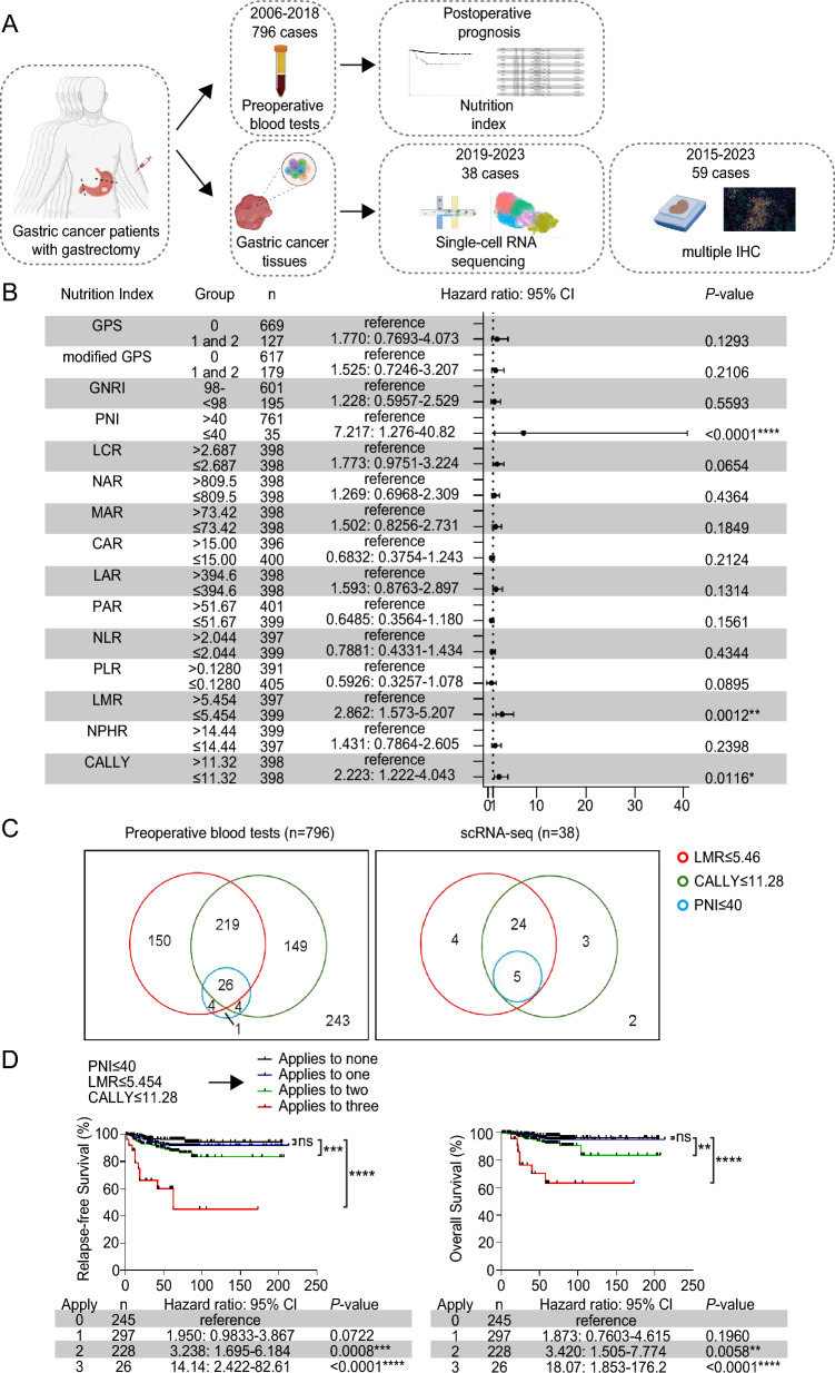
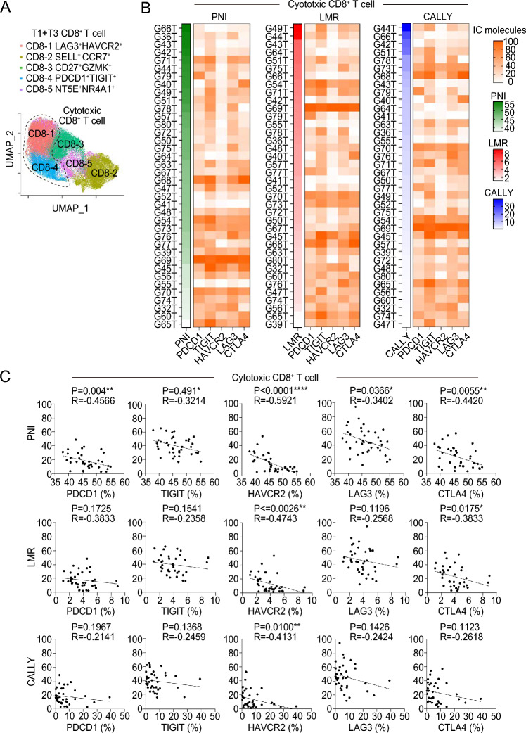
Materials and methods: We retrospectively analyzed clinical data from 796 GC patients who underwent radical gastrectomy to identify which previously reported nutritional index had the greatest impact on prognosis. Single-cell RNA sequencing was performed on 38 GC tissues, and multiplex immunofluorescence staining was conducted on 59 GC tissues to evaluate the relationship between nutritional indices and IC molecule expression in cytotoxic CD8-positive T cells.
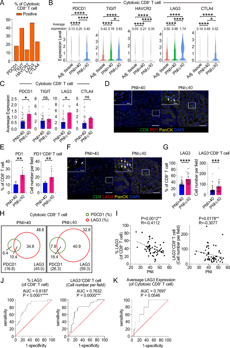
Results: A low preoperative PNI was identified as the strongest predictor of poor prognosis among the nutritional indices in GC patients. The expression of not only PDCD1 (encoding PD1) but also LAG3 in cytotoxic CD8-positive T cells was significantly higher in GC with low PNI compared to those with high PNI. Among cytotoxic CD8-positive T cells, the proportion of LAG3-positive cells was greater than that of PDCD1-positive cells, particularly in GC with low PNI, and most LAG3-positive cells did not co-express PDCD1. Additionally, the expression of MHC class II, a ligand for LAG3, was higher in GC cells with high levels of epithelial-mesenchymal transition-related molecules in GC with low PNI compared to those with high PNI.
Conclusions: PNI can reflect LAG3 expression in cytotoxic CD8-positive T cells and MHC class II expression in GC cells.
期刊介绍:
Cancer Immunology, Immunotherapy has the basic aim of keeping readers informed of the latest research results in the fields of oncology and immunology. As knowledge expands, the scope of the journal has broadened to include more of the progress being made in the areas of biology concerned with biological response modifiers. This helps keep readers up to date on the latest advances in our understanding of tumor-host interactions.
The journal publishes short editorials including "position papers," general reviews, original articles, and short communications, providing a forum for the most current experimental and clinical advances in tumor immunology.
 求助内容:
求助内容: 应助结果提醒方式:


