M Campagnolo, M Puthenparampil, A Emmi, L Weis, E Basili, V Mauceri, A Miscioscia, M Carecchio, A Guerra, V Misenti, C Fogliano, P Gallo, A Antonini
{"title":"光学相干断层扫描显示α-突触核蛋白病的视网膜结构异常:来自Padua-CESNE队列的见解。","authors":"M Campagnolo, M Puthenparampil, A Emmi, L Weis, E Basili, V Mauceri, A Miscioscia, M Carecchio, A Guerra, V Misenti, C Fogliano, P Gallo, A Antonini","doi":"10.1007/s00702-025-02918-y","DOIUrl":null,"url":null,"abstract":"<p><p>The complexity of α-synucleinopathies, namely Parkinson's disease (PD) and multiple system atrophy (MSA), calls for the adoption a multimodal approach integrating biological, morphological, and functional data. Phosphorylated α-synuclein (α-syn) detection in bodily fluids and tissues such as the skin helps provide biological characterization of the disease, but specific and accessible biomarkers are not available yet. The aim of this study was to define the role of Optical Coherence Tomography (OCT, a minimally invasive retinal imaging technique) patterns as possible biomarkers in the early stages of α-synucleinopathies, also supporting the differential diagnosis. Thirty-five (23 PD, 12 MSA), clinically, biologically and genetically characterized patients included in the PADUA-CESNE (Centro Studi per la Neurodegenerazione) cohort underwent OCT. A significant atrophy in the inferior, superior and temporal regions of the Retinal Nerve Fiber Layer (RNFL) and in the inner nuclear layer (INL) were observed in PD compared to controls, differently from MSA. Hyperreflective foci (HRF) counts were elevated across all retinal layers in all patients with PD exhibiting significantly higher numbers, suggesting microglial activation and greater retinal damage. Further research regarding OCT patterns in PD and MSA may consolidate the role of specific features, such as INL abnormalities and different HRF counts, in supporting the diagnosis and differential diagnosis in α-synucleinopathies. In light of the availability of potentially disease-modifying therapies, studies should focus on newly diagnosed patients, also undergoing thorough clinical, biological and genetic characterization.</p>","PeriodicalId":16579,"journal":{"name":"Journal of Neural Transmission","volume":" ","pages":"1013-1022"},"PeriodicalIF":4.0000,"publicationDate":"2025-07-01","publicationTypes":"Journal Article","fieldsOfStudy":null,"isOpenAccess":false,"openAccessPdf":"https://www.ncbi.nlm.nih.gov/pmc/articles/PMC12208970/pdf/","citationCount":"0","resultStr":"{\"title\":\"Optical coherence tomography reveals retinal structural abnormalities in α-synucleinopathies: insights from the Padua-CESNE cohort.\",\"authors\":\"M Campagnolo, M Puthenparampil, A Emmi, L Weis, E Basili, V Mauceri, A Miscioscia, M Carecchio, A Guerra, V Misenti, C Fogliano, P Gallo, A Antonini\",\"doi\":\"10.1007/s00702-025-02918-y\",\"DOIUrl\":null,\"url\":null,\"abstract\":\"<p><p>The complexity of α-synucleinopathies, namely Parkinson's disease (PD) and multiple system atrophy (MSA), calls for the adoption a multimodal approach integrating biological, morphological, and functional data. Phosphorylated α-synuclein (α-syn) detection in bodily fluids and tissues such as the skin helps provide biological characterization of the disease, but specific and accessible biomarkers are not available yet. The aim of this study was to define the role of Optical Coherence Tomography (OCT, a minimally invasive retinal imaging technique) patterns as possible biomarkers in the early stages of α-synucleinopathies, also supporting the differential diagnosis. Thirty-five (23 PD, 12 MSA), clinically, biologically and genetically characterized patients included in the PADUA-CESNE (Centro Studi per la Neurodegenerazione) cohort underwent OCT. A significant atrophy in the inferior, superior and temporal regions of the Retinal Nerve Fiber Layer (RNFL) and in the inner nuclear layer (INL) were observed in PD compared to controls, differently from MSA. Hyperreflective foci (HRF) counts were elevated across all retinal layers in all patients with PD exhibiting significantly higher numbers, suggesting microglial activation and greater retinal damage. Further research regarding OCT patterns in PD and MSA may consolidate the role of specific features, such as INL abnormalities and different HRF counts, in supporting the diagnosis and differential diagnosis in α-synucleinopathies. In light of the availability of potentially disease-modifying therapies, studies should focus on newly diagnosed patients, also undergoing thorough clinical, biological and genetic characterization.</p>\",\"PeriodicalId\":16579,\"journal\":{\"name\":\"Journal of Neural Transmission\",\"volume\":\" \",\"pages\":\"1013-1022\"},\"PeriodicalIF\":4.0000,\"publicationDate\":\"2025-07-01\",\"publicationTypes\":\"Journal Article\",\"fieldsOfStudy\":null,\"isOpenAccess\":false,\"openAccessPdf\":\"https://www.ncbi.nlm.nih.gov/pmc/articles/PMC12208970/pdf/\",\"citationCount\":\"0\",\"resultStr\":null,\"platform\":\"Semanticscholar\",\"paperid\":null,\"PeriodicalName\":\"Journal of Neural Transmission\",\"FirstCategoryId\":\"3\",\"ListUrlMain\":\"https://doi.org/10.1007/s00702-025-02918-y\",\"RegionNum\":4,\"RegionCategory\":\"医学\",\"ArticlePicture\":[],\"TitleCN\":null,\"AbstractTextCN\":null,\"PMCID\":null,\"EPubDate\":\"2025/4/15 0:00:00\",\"PubModel\":\"Epub\",\"JCR\":\"Q2\",\"JCRName\":\"CLINICAL NEUROLOGY\",\"Score\":null,\"Total\":0}","platform":"Semanticscholar","paperid":null,"PeriodicalName":"Journal of Neural Transmission","FirstCategoryId":"3","ListUrlMain":"https://doi.org/10.1007/s00702-025-02918-y","RegionNum":4,"RegionCategory":"医学","ArticlePicture":[],"TitleCN":null,"AbstractTextCN":null,"PMCID":null,"EPubDate":"2025/4/15 0:00:00","PubModel":"Epub","JCR":"Q2","JCRName":"CLINICAL NEUROLOGY","Score":null,"Total":0}
引用次数: 0
摘要
α-突触核蛋白病,即帕金森病(PD)和多系统萎缩(MSA)的复杂性要求采用多模式方法整合生物学,形态学和功能数据。在体液和皮肤等组织中检测磷酸化α-突触核蛋白(α-syn)有助于提供该疾病的生物学特征,但目前还没有特异性和可获得的生物标志物。本研究的目的是确定光学相干断层扫描(OCT,一种微创视网膜成像技术)模式在α-突触核蛋白病早期可能的生物标志物的作用,并支持鉴别诊断。在PADUA-CESNE (Centro Studi per la Neurodegenerazione)队列中,35例(23例PD, 12例MSA)具有临床、生物学和遗传学特征的患者接受了oct治疗。与对照组相比,PD患者的视网膜神经纤维层(RNFL)的下、上、颞区和内核层(INL)明显萎缩,这与MSA不同。所有PD患者视网膜各层的高反射灶(Hyperreflective foci, HRF)计数均升高,显示出明显较高的数字,提示小胶质细胞激活和更大的视网膜损伤。进一步研究PD和MSA的OCT模式可能会巩固特定特征(如INL异常和不同HRF计数)在支持α-突触核蛋白病的诊断和鉴别诊断中的作用。鉴于有可能改善疾病的治疗方法,研究应集中在新诊断的患者身上,也要进行彻底的临床、生物学和遗传学鉴定。
Optical coherence tomography reveals retinal structural abnormalities in α-synucleinopathies: insights from the Padua-CESNE cohort.
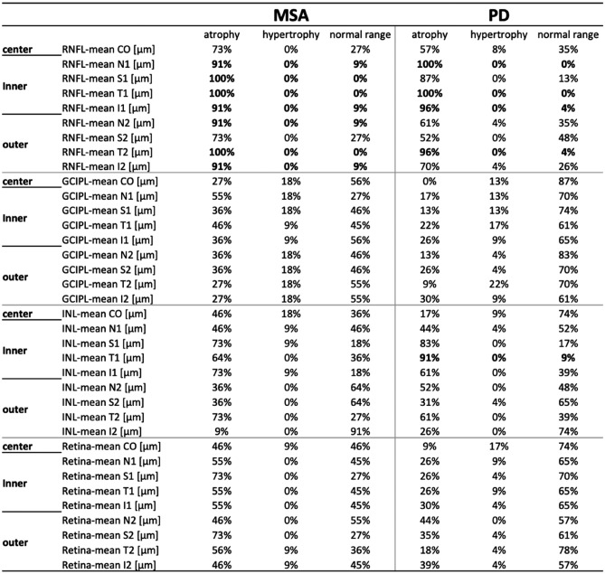
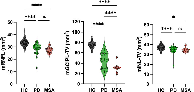
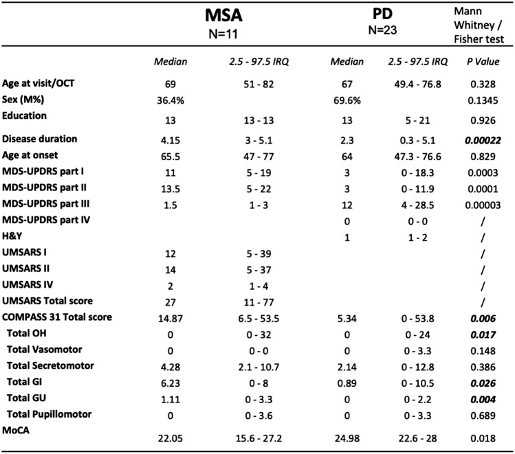
The complexity of α-synucleinopathies, namely Parkinson's disease (PD) and multiple system atrophy (MSA), calls for the adoption a multimodal approach integrating biological, morphological, and functional data. Phosphorylated α-synuclein (α-syn) detection in bodily fluids and tissues such as the skin helps provide biological characterization of the disease, but specific and accessible biomarkers are not available yet. The aim of this study was to define the role of Optical Coherence Tomography (OCT, a minimally invasive retinal imaging technique) patterns as possible biomarkers in the early stages of α-synucleinopathies, also supporting the differential diagnosis. Thirty-five (23 PD, 12 MSA), clinically, biologically and genetically characterized patients included in the PADUA-CESNE (Centro Studi per la Neurodegenerazione) cohort underwent OCT. A significant atrophy in the inferior, superior and temporal regions of the Retinal Nerve Fiber Layer (RNFL) and in the inner nuclear layer (INL) were observed in PD compared to controls, differently from MSA. Hyperreflective foci (HRF) counts were elevated across all retinal layers in all patients with PD exhibiting significantly higher numbers, suggesting microglial activation and greater retinal damage. Further research regarding OCT patterns in PD and MSA may consolidate the role of specific features, such as INL abnormalities and different HRF counts, in supporting the diagnosis and differential diagnosis in α-synucleinopathies. In light of the availability of potentially disease-modifying therapies, studies should focus on newly diagnosed patients, also undergoing thorough clinical, biological and genetic characterization.
期刊介绍:
The investigation of basic mechanisms involved in the pathogenesis of neurological and psychiatric disorders has undoubtedly deepened our knowledge of these types of disorders. The impact of basic neurosciences on the understanding of the pathophysiology of the brain will further increase due to important developments such as the emergence of more specific psychoactive compounds and new technologies.
The Journal of Neural Transmission aims to establish an interface between basic sciences and clinical neurology and psychiatry. It intends to put a special emphasis on translational publications of the newest developments in the field from all disciplines of the neural sciences that relate to a better understanding and treatment of neurological and psychiatric disorders.
 求助内容:
求助内容: 应助结果提醒方式:


