{"title":"蜂胶水溶液提取物降低乳牙粪肠球菌数量的体外研究。","authors":"Majd Refaay, Mhd Bashier Almonakel, Samar Alsalameh, Ibraheem Hawary, Yasser Alsayed Tolibah","doi":"10.1155/ijod/7629615","DOIUrl":null,"url":null,"abstract":"<p><p><b>Objective:</b> To evaluate the efficacy of 11% aqueous propolis extract in eliminating <i>Enterococcus faecalis</i> in necrotic pulp canals of primary anterior teeth compared to 2.5% sodium hypochlorite. <b>Materials</b> and Methods: <i>E. faecalis</i> were isolated from necrotic primary anterior teeth with periapical lesions, cultured, and incubated using paper points. The research sample comprised 30 extracted single-rooted necrotic primary anterior teeth, divided equally into two groups according to the irrigants used. Access cavities were prepared, and working lengths were determined. Afterward, canals were shaped using K-files, contaminated with <i>E. faecalis</i>, and placed in an incubator for a week. Initial microbial swabs were taken, and then each canal was irrigated with either 3 mL of a hand-made 11% aqueous propolis extract or 3 mL of 2.5% sodium hypochlorite for 5 min. Postirrigation microbial swabs were taken, cultured on blood agar plates, and incubated at 37°C for 48 h, followed by colony counts. Statistical tests included the paired sample <i>T</i>-test, Wilcoxon signed ranks, and Mann-Whitney <i>U</i> tests. The significance level was set at <i>α</i> = 0.05. <b>Results:</b> In total, 11% aqueous propolis extract contributed to a 61.8% reduction in <i>E. faecalis</i> (<i>p</i> < 0.001), while 2.5% sodium hypochlorite contributed to an 84.1% reduction (<i>p</i> < 0.001). The average change in logarithmic values in the sodium hypochlorite group was more significant than in the propolis group (<i>p</i>=0.002). <b>Conclusion:</b> In total, 11% aqueous propolis extract is antimicrobial against <i>E. faecalis</i>. However, its efficacy was less than 2.5% sodium hypochlorite.</p>","PeriodicalId":13947,"journal":{"name":"International Journal of Dentistry","volume":"2025 ","pages":"7629615"},"PeriodicalIF":2.2000,"publicationDate":"2025-04-04","publicationTypes":"Journal Article","fieldsOfStudy":null,"isOpenAccess":false,"openAccessPdf":"https://www.ncbi.nlm.nih.gov/pmc/articles/PMC11991864/pdf/","citationCount":"0","resultStr":"{\"title\":\"The Effectiveness of Aqueous Propolis Extract in Reducing the <i>Enterococcus faecalis</i> Count in Primary Teeth: An In Vitro Study.\",\"authors\":\"Majd Refaay, Mhd Bashier Almonakel, Samar Alsalameh, Ibraheem Hawary, Yasser Alsayed Tolibah\",\"doi\":\"10.1155/ijod/7629615\",\"DOIUrl\":null,\"url\":null,\"abstract\":\"<p><p><b>Objective:</b> To evaluate the efficacy of 11% aqueous propolis extract in eliminating <i>Enterococcus faecalis</i> in necrotic pulp canals of primary anterior teeth compared to 2.5% sodium hypochlorite. <b>Materials</b> and Methods: <i>E. faecalis</i> were isolated from necrotic primary anterior teeth with periapical lesions, cultured, and incubated using paper points. The research sample comprised 30 extracted single-rooted necrotic primary anterior teeth, divided equally into two groups according to the irrigants used. Access cavities were prepared, and working lengths were determined. Afterward, canals were shaped using K-files, contaminated with <i>E. faecalis</i>, and placed in an incubator for a week. Initial microbial swabs were taken, and then each canal was irrigated with either 3 mL of a hand-made 11% aqueous propolis extract or 3 mL of 2.5% sodium hypochlorite for 5 min. Postirrigation microbial swabs were taken, cultured on blood agar plates, and incubated at 37°C for 48 h, followed by colony counts. Statistical tests included the paired sample <i>T</i>-test, Wilcoxon signed ranks, and Mann-Whitney <i>U</i> tests. The significance level was set at <i>α</i> = 0.05. <b>Results:</b> In total, 11% aqueous propolis extract contributed to a 61.8% reduction in <i>E. faecalis</i> (<i>p</i> < 0.001), while 2.5% sodium hypochlorite contributed to an 84.1% reduction (<i>p</i> < 0.001). The average change in logarithmic values in the sodium hypochlorite group was more significant than in the propolis group (<i>p</i>=0.002). <b>Conclusion:</b> In total, 11% aqueous propolis extract is antimicrobial against <i>E. faecalis</i>. However, its efficacy was less than 2.5% sodium hypochlorite.</p>\",\"PeriodicalId\":13947,\"journal\":{\"name\":\"International Journal of Dentistry\",\"volume\":\"2025 \",\"pages\":\"7629615\"},\"PeriodicalIF\":2.2000,\"publicationDate\":\"2025-04-04\",\"publicationTypes\":\"Journal Article\",\"fieldsOfStudy\":null,\"isOpenAccess\":false,\"openAccessPdf\":\"https://www.ncbi.nlm.nih.gov/pmc/articles/PMC11991864/pdf/\",\"citationCount\":\"0\",\"resultStr\":null,\"platform\":\"Semanticscholar\",\"paperid\":null,\"PeriodicalName\":\"International Journal of Dentistry\",\"FirstCategoryId\":\"1085\",\"ListUrlMain\":\"https://doi.org/10.1155/ijod/7629615\",\"RegionNum\":0,\"RegionCategory\":null,\"ArticlePicture\":[],\"TitleCN\":null,\"AbstractTextCN\":null,\"PMCID\":null,\"EPubDate\":\"2025/1/1 0:00:00\",\"PubModel\":\"eCollection\",\"JCR\":\"Q2\",\"JCRName\":\"DENTISTRY, ORAL SURGERY & MEDICINE\",\"Score\":null,\"Total\":0}","platform":"Semanticscholar","paperid":null,"PeriodicalName":"International Journal of Dentistry","FirstCategoryId":"1085","ListUrlMain":"https://doi.org/10.1155/ijod/7629615","RegionNum":0,"RegionCategory":null,"ArticlePicture":[],"TitleCN":null,"AbstractTextCN":null,"PMCID":null,"EPubDate":"2025/1/1 0:00:00","PubModel":"eCollection","JCR":"Q2","JCRName":"DENTISTRY, ORAL SURGERY & MEDICINE","Score":null,"Total":0}
The Effectiveness of Aqueous Propolis Extract in Reducing the Enterococcus faecalis Count in Primary Teeth: An In Vitro Study.
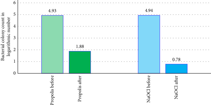
Objective: To evaluate the efficacy of 11% aqueous propolis extract in eliminating Enterococcus faecalis in necrotic pulp canals of primary anterior teeth compared to 2.5% sodium hypochlorite. Materials and Methods: E. faecalis were isolated from necrotic primary anterior teeth with periapical lesions, cultured, and incubated using paper points. The research sample comprised 30 extracted single-rooted necrotic primary anterior teeth, divided equally into two groups according to the irrigants used. Access cavities were prepared, and working lengths were determined. Afterward, canals were shaped using K-files, contaminated with E. faecalis, and placed in an incubator for a week. Initial microbial swabs were taken, and then each canal was irrigated with either 3 mL of a hand-made 11% aqueous propolis extract or 3 mL of 2.5% sodium hypochlorite for 5 min. Postirrigation microbial swabs were taken, cultured on blood agar plates, and incubated at 37°C for 48 h, followed by colony counts. Statistical tests included the paired sample T-test, Wilcoxon signed ranks, and Mann-Whitney U tests. The significance level was set at α = 0.05. Results: In total, 11% aqueous propolis extract contributed to a 61.8% reduction in E. faecalis (p < 0.001), while 2.5% sodium hypochlorite contributed to an 84.1% reduction (p < 0.001). The average change in logarithmic values in the sodium hypochlorite group was more significant than in the propolis group (p=0.002). Conclusion: In total, 11% aqueous propolis extract is antimicrobial against E. faecalis. However, its efficacy was less than 2.5% sodium hypochlorite.

 求助内容:
求助内容: 应助结果提醒方式:


