{"title":"利用H215O和正电子发射断层扫描缩短脑血显现时间成像计算时间的试验。","authors":"Takuya Kobata, Takashi Norikane, Mitsumasa Murao, Yuri Manabe, Yuka Yamamoto, Katsuya Mitamura, Tetsuhiro Hatakeyama, Keisuke Miyake, Yoshihiro Nishiyama, Nobuyuki Kudomi","doi":"10.1007/s12149-025-02054-3","DOIUrl":null,"url":null,"abstract":"<div><h3>Objective</h3><p>Delayed appearance of blood in the brain may be a pathophysiological indicator of stenosis or occlusion. Image computation for blood appearance generally requires considerable time. Therefore, in this study, we aimed to shorten the computation time using several algorithms and tested their accuracy and precision using examination data and simulations, as well as the computation time.</p><h3>Methods</h3><p>We retrospectively analyzed the images of patients with suspected cerebrovascular disorders who underwent PET study with <sup>15</sup>O-labeled tracers. The blood appearance time images were computed by fitting a stepwise time-shifted tissue curve and applying a single-tissue compartment model with several modes of fixing or not fixing the washout rate and/or blood volume terms. The appearance times in images for these modes were compared with the time obtained by ROI-based non-linear fitting in several brain regions. The effects of noise and parameter fixation are assessed using a simulation study.</p><h3>Results</h3><p>The computation time was 28.2 ± 6.2 min, 16.6 ± 3.9 min, and 2.6 ± 1.1 min for modes without fixing, with fixing blood volume, and washout rate, respectively. The mean difference in the appearance time against ROI-based non-linear fitting was less than 1 s with a standard deviation (SD) of approximately 2.5 s for those modes. The images obtained were similar for all three modes. The simulation showed that SD on the estimated appearance times were acceptable, namely < 1.5 s, for these modes.</p><h3>Conclusion</h3><p>This study suggests the possibility of visualizing appearance time images in the brain with a reasonable computation time of approximately 2.5 min at the minimum and 30 min at the most.</p></div>","PeriodicalId":8007,"journal":{"name":"Annals of Nuclear Medicine","volume":"39 8","pages":"823 - 832"},"PeriodicalIF":2.5000,"publicationDate":"2025-04-29","publicationTypes":"Journal Article","fieldsOfStudy":null,"isOpenAccess":false,"openAccessPdf":"","citationCount":"0","resultStr":"{\"title\":\"Test for shortening the computation time for imaging of appearance time for cerebral blood using H215O and positron emission tomography\",\"authors\":\"Takuya Kobata, Takashi Norikane, Mitsumasa Murao, Yuri Manabe, Yuka Yamamoto, Katsuya Mitamura, Tetsuhiro Hatakeyama, Keisuke Miyake, Yoshihiro Nishiyama, Nobuyuki Kudomi\",\"doi\":\"10.1007/s12149-025-02054-3\",\"DOIUrl\":null,\"url\":null,\"abstract\":\"<div><h3>Objective</h3><p>Delayed appearance of blood in the brain may be a pathophysiological indicator of stenosis or occlusion. Image computation for blood appearance generally requires considerable time. Therefore, in this study, we aimed to shorten the computation time using several algorithms and tested their accuracy and precision using examination data and simulations, as well as the computation time.</p><h3>Methods</h3><p>We retrospectively analyzed the images of patients with suspected cerebrovascular disorders who underwent PET study with <sup>15</sup>O-labeled tracers. The blood appearance time images were computed by fitting a stepwise time-shifted tissue curve and applying a single-tissue compartment model with several modes of fixing or not fixing the washout rate and/or blood volume terms. The appearance times in images for these modes were compared with the time obtained by ROI-based non-linear fitting in several brain regions. The effects of noise and parameter fixation are assessed using a simulation study.</p><h3>Results</h3><p>The computation time was 28.2 ± 6.2 min, 16.6 ± 3.9 min, and 2.6 ± 1.1 min for modes without fixing, with fixing blood volume, and washout rate, respectively. The mean difference in the appearance time against ROI-based non-linear fitting was less than 1 s with a standard deviation (SD) of approximately 2.5 s for those modes. The images obtained were similar for all three modes. The simulation showed that SD on the estimated appearance times were acceptable, namely < 1.5 s, for these modes.</p><h3>Conclusion</h3><p>This study suggests the possibility of visualizing appearance time images in the brain with a reasonable computation time of approximately 2.5 min at the minimum and 30 min at the most.</p></div>\",\"PeriodicalId\":8007,\"journal\":{\"name\":\"Annals of Nuclear Medicine\",\"volume\":\"39 8\",\"pages\":\"823 - 832\"},\"PeriodicalIF\":2.5000,\"publicationDate\":\"2025-04-29\",\"publicationTypes\":\"Journal Article\",\"fieldsOfStudy\":null,\"isOpenAccess\":false,\"openAccessPdf\":\"\",\"citationCount\":\"0\",\"resultStr\":null,\"platform\":\"Semanticscholar\",\"paperid\":null,\"PeriodicalName\":\"Annals of Nuclear Medicine\",\"FirstCategoryId\":\"3\",\"ListUrlMain\":\"https://link.springer.com/article/10.1007/s12149-025-02054-3\",\"RegionNum\":4,\"RegionCategory\":\"医学\",\"ArticlePicture\":[],\"TitleCN\":null,\"AbstractTextCN\":null,\"PMCID\":null,\"EPubDate\":\"\",\"PubModel\":\"\",\"JCR\":\"Q2\",\"JCRName\":\"RADIOLOGY, NUCLEAR MEDICINE & MEDICAL IMAGING\",\"Score\":null,\"Total\":0}","platform":"Semanticscholar","paperid":null,"PeriodicalName":"Annals of Nuclear Medicine","FirstCategoryId":"3","ListUrlMain":"https://link.springer.com/article/10.1007/s12149-025-02054-3","RegionNum":4,"RegionCategory":"医学","ArticlePicture":[],"TitleCN":null,"AbstractTextCN":null,"PMCID":null,"EPubDate":"","PubModel":"","JCR":"Q2","JCRName":"RADIOLOGY, NUCLEAR MEDICINE & MEDICAL IMAGING","Score":null,"Total":0}
Test for shortening the computation time for imaging of appearance time for cerebral blood using H215O and positron emission tomography
Objective
Delayed appearance of blood in the brain may be a pathophysiological indicator of stenosis or occlusion. Image computation for blood appearance generally requires considerable time. Therefore, in this study, we aimed to shorten the computation time using several algorithms and tested their accuracy and precision using examination data and simulations, as well as the computation time.
Methods
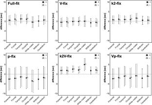
We retrospectively analyzed the images of patients with suspected cerebrovascular disorders who underwent PET study with 15O-labeled tracers. The blood appearance time images were computed by fitting a stepwise time-shifted tissue curve and applying a single-tissue compartment model with several modes of fixing or not fixing the washout rate and/or blood volume terms. The appearance times in images for these modes were compared with the time obtained by ROI-based non-linear fitting in several brain regions. The effects of noise and parameter fixation are assessed using a simulation study.
Results
The computation time was 28.2 ± 6.2 min, 16.6 ± 3.9 min, and 2.6 ± 1.1 min for modes without fixing, with fixing blood volume, and washout rate, respectively. The mean difference in the appearance time against ROI-based non-linear fitting was less than 1 s with a standard deviation (SD) of approximately 2.5 s for those modes. The images obtained were similar for all three modes. The simulation showed that SD on the estimated appearance times were acceptable, namely < 1.5 s, for these modes.
Conclusion
This study suggests the possibility of visualizing appearance time images in the brain with a reasonable computation time of approximately 2.5 min at the minimum and 30 min at the most.
期刊介绍:
Annals of Nuclear Medicine is an official journal of the Japanese Society of Nuclear Medicine. It develops the appropriate application of radioactive substances and stable nuclides in the field of medicine.
The journal promotes the exchange of ideas and information and research in nuclear medicine and includes the medical application of radionuclides and related subjects. It presents original articles, short communications, reviews and letters to the editor.

 求助内容:
求助内容: 应助结果提醒方式:


