{"title":"原发性皮肤黑色素瘤的脉络膜转移:1例报告。","authors":"Teele Palumaa, Artur Klett","doi":"10.1159/000544879","DOIUrl":null,"url":null,"abstract":"<p><strong>Introduction: </strong>Choroidal metastases most often originate from breast tumors in females and lung cancer in males. Primary cutaneous tumors rarely metastasize to the uveal tract.</p><p><strong>Case presentation: </strong>Here, we present a rare case of a 47-year-old female patient who was clinically diagnosed with a presumed choroidal metastasis of primary cutaneous melanoma. The patient presented with complaints of decreased vision in her right eye. Her best corrected visual acuity (BCVA) was 0.7 decimal in the right eye and 1.0 in the left eye. Examination revealed a pigmented choroidal lesion in the parafoveal region with a prominence of 2.9 mm, orange pigment on the surface, and subretinal fluid in its projection. She had no history of active malignancy, but at the age of 36, a localized stage IB cutaneous melanoma was removed from her back. Yearly follow-up visits at the dermatologist showed no evidence of active disease. Upon diagnosis of a choroidal tumor, the patient underwent brachytherapy with a ruthenium-106 plaque in the right eye. Follow-up at the oncologist revealed a widespread disease with metastases in distant lymph nodes, liver, lung, pancreas, and brain, an uncommon pattern for primary choroidal melanomas, resembling rather the metastasis pattern of primary cutaneous melanoma. The patient was started on systemic therapy against metastatic cutaneous melanoma. At 21 months after brachytherapy and 19 months after the initiation of systemic anticancer therapy, the patient's BCVA in the right eye returned to 1.0 decimal, the choroidal lesion reduced in size, and subretinal fluid receded. Two years after the initial presentation, all metastases were stable or decreased in size.</p><p><strong>Conclusion: </strong>This case highlights the possibility of a choroidal metastasis of cutaneous melanoma more than a decade after the first presentation of the disease and highlights the effectiveness of combined brachytherapy and systemic anticancer therapy in managing the disease.</p>","PeriodicalId":9635,"journal":{"name":"Case Reports in Ophthalmology","volume":"16 1","pages":"267-273"},"PeriodicalIF":0.6000,"publicationDate":"2025-03-24","publicationTypes":"Journal Article","fieldsOfStudy":null,"isOpenAccess":false,"openAccessPdf":"https://www.ncbi.nlm.nih.gov/pmc/articles/PMC12002729/pdf/","citationCount":"0","resultStr":"{\"title\":\"Presumed Choroidal Metastasis of Primary Cutaneous Melanoma: A Case Report.\",\"authors\":\"Teele Palumaa, Artur Klett\",\"doi\":\"10.1159/000544879\",\"DOIUrl\":null,\"url\":null,\"abstract\":\"<p><strong>Introduction: </strong>Choroidal metastases most often originate from breast tumors in females and lung cancer in males. Primary cutaneous tumors rarely metastasize to the uveal tract.</p><p><strong>Case presentation: </strong>Here, we present a rare case of a 47-year-old female patient who was clinically diagnosed with a presumed choroidal metastasis of primary cutaneous melanoma. The patient presented with complaints of decreased vision in her right eye. Her best corrected visual acuity (BCVA) was 0.7 decimal in the right eye and 1.0 in the left eye. Examination revealed a pigmented choroidal lesion in the parafoveal region with a prominence of 2.9 mm, orange pigment on the surface, and subretinal fluid in its projection. She had no history of active malignancy, but at the age of 36, a localized stage IB cutaneous melanoma was removed from her back. Yearly follow-up visits at the dermatologist showed no evidence of active disease. Upon diagnosis of a choroidal tumor, the patient underwent brachytherapy with a ruthenium-106 plaque in the right eye. Follow-up at the oncologist revealed a widespread disease with metastases in distant lymph nodes, liver, lung, pancreas, and brain, an uncommon pattern for primary choroidal melanomas, resembling rather the metastasis pattern of primary cutaneous melanoma. The patient was started on systemic therapy against metastatic cutaneous melanoma. At 21 months after brachytherapy and 19 months after the initiation of systemic anticancer therapy, the patient's BCVA in the right eye returned to 1.0 decimal, the choroidal lesion reduced in size, and subretinal fluid receded. Two years after the initial presentation, all metastases were stable or decreased in size.</p><p><strong>Conclusion: </strong>This case highlights the possibility of a choroidal metastasis of cutaneous melanoma more than a decade after the first presentation of the disease and highlights the effectiveness of combined brachytherapy and systemic anticancer therapy in managing the disease.</p>\",\"PeriodicalId\":9635,\"journal\":{\"name\":\"Case Reports in Ophthalmology\",\"volume\":\"16 1\",\"pages\":\"267-273\"},\"PeriodicalIF\":0.6000,\"publicationDate\":\"2025-03-24\",\"publicationTypes\":\"Journal Article\",\"fieldsOfStudy\":null,\"isOpenAccess\":false,\"openAccessPdf\":\"https://www.ncbi.nlm.nih.gov/pmc/articles/PMC12002729/pdf/\",\"citationCount\":\"0\",\"resultStr\":null,\"platform\":\"Semanticscholar\",\"paperid\":null,\"PeriodicalName\":\"Case Reports in Ophthalmology\",\"FirstCategoryId\":\"1085\",\"ListUrlMain\":\"https://doi.org/10.1159/000544879\",\"RegionNum\":0,\"RegionCategory\":null,\"ArticlePicture\":[],\"TitleCN\":null,\"AbstractTextCN\":null,\"PMCID\":null,\"EPubDate\":\"2025/1/1 0:00:00\",\"PubModel\":\"eCollection\",\"JCR\":\"Q4\",\"JCRName\":\"OPHTHALMOLOGY\",\"Score\":null,\"Total\":0}","platform":"Semanticscholar","paperid":null,"PeriodicalName":"Case Reports in Ophthalmology","FirstCategoryId":"1085","ListUrlMain":"https://doi.org/10.1159/000544879","RegionNum":0,"RegionCategory":null,"ArticlePicture":[],"TitleCN":null,"AbstractTextCN":null,"PMCID":null,"EPubDate":"2025/1/1 0:00:00","PubModel":"eCollection","JCR":"Q4","JCRName":"OPHTHALMOLOGY","Score":null,"Total":0}
Presumed Choroidal Metastasis of Primary Cutaneous Melanoma: A Case Report.
Introduction: Choroidal metastases most often originate from breast tumors in females and lung cancer in males. Primary cutaneous tumors rarely metastasize to the uveal tract.
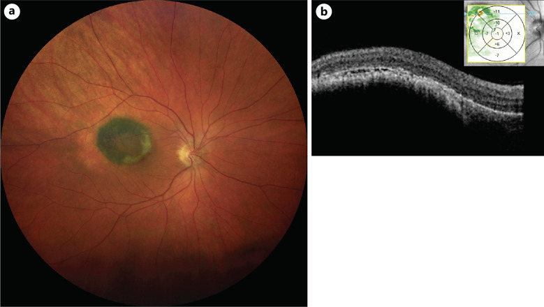
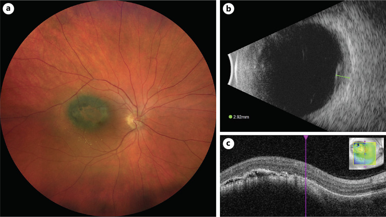
Case presentation: Here, we present a rare case of a 47-year-old female patient who was clinically diagnosed with a presumed choroidal metastasis of primary cutaneous melanoma. The patient presented with complaints of decreased vision in her right eye. Her best corrected visual acuity (BCVA) was 0.7 decimal in the right eye and 1.0 in the left eye. Examination revealed a pigmented choroidal lesion in the parafoveal region with a prominence of 2.9 mm, orange pigment on the surface, and subretinal fluid in its projection. She had no history of active malignancy, but at the age of 36, a localized stage IB cutaneous melanoma was removed from her back. Yearly follow-up visits at the dermatologist showed no evidence of active disease. Upon diagnosis of a choroidal tumor, the patient underwent brachytherapy with a ruthenium-106 plaque in the right eye. Follow-up at the oncologist revealed a widespread disease with metastases in distant lymph nodes, liver, lung, pancreas, and brain, an uncommon pattern for primary choroidal melanomas, resembling rather the metastasis pattern of primary cutaneous melanoma. The patient was started on systemic therapy against metastatic cutaneous melanoma. At 21 months after brachytherapy and 19 months after the initiation of systemic anticancer therapy, the patient's BCVA in the right eye returned to 1.0 decimal, the choroidal lesion reduced in size, and subretinal fluid receded. Two years after the initial presentation, all metastases were stable or decreased in size.
Conclusion: This case highlights the possibility of a choroidal metastasis of cutaneous melanoma more than a decade after the first presentation of the disease and highlights the effectiveness of combined brachytherapy and systemic anticancer therapy in managing the disease.
期刊介绍:
This peer-reviewed online-only journal publishes original case reports covering the entire spectrum of ophthalmology, including prevention, diagnosis, treatment, toxicities of therapy, supportive care, quality-of-life, and survivorship issues. The submission of negative results is strongly encouraged. The journal will also accept case reports dealing with the use of novel technologies, both in the arena of diagnosis and treatment. Supplementary material is welcomed. The intent of the journal is to provide clinicians and researchers with a tool to disseminate their personal experiences to a wider public as well as to review interesting cases encountered by colleagues all over the world. Universally used terms can be searched across the entire growing collection of case reports, further facilitating the retrieval of specific information. Following the open access principle, the entire contents can be retrieved at no charge, guaranteeing easy access to this valuable source of anecdotal information at all times.

 求助内容:
求助内容: 应助结果提醒方式:


