{"title":"猫单侧肺静脉狭窄的死前诊断1例。","authors":"Takuma Aoki, Takashi Miyamoto, Kota Kizaki, Asuka Ueshima, Kentaro Iwasaki, Takuya Kusaka, Haruko Terui","doi":"10.1186/s13028-025-00803-y","DOIUrl":null,"url":null,"abstract":"<p><strong>Background: </strong>Pulmonary hypertension (PH) detection in cats may be challenging. Pulmonary venous stenosis (PVS) is rare in cats and can lead to PH. The only reported PVS case received a post-mortem diagnosis. Imaging during the cat's lifetime established the diagnosis in this case.</p><p><strong>Case presentation: </strong>A 2 year-old Norwegian Forest cat was diagnosed with pulmonary oedema and PH secondary to cor triatriatum sinister (CTS) and showed improved breathing following two subcutaneous furosemide treatments, 1 and 2 mg/kg, during an overnight stay at the referral veterinary hospital. Sildenafil alone (0.69 mg/kg, PO, BID) was prescribed post-discharge to address PH without diuretics. Post-discharge from the referral veterinary hospital, collapse and pre-syncope were suspected to be due to PH. Consequently, sildenafil was titrated weekly, starting at 1.09 mg/kg BID and increasing to 1.63 mg/kg BID. Pre-syncope and collapse resolved, and pulmonary opacities reduced considerably, although concerns remained that increased pulmonary blood flow to suspected CTS from sildenafil might worsen cardiogenic pulmonary oedema. The patient was also treated with rivaroxaban (2.5 mg/head, SID), considering the increased risk of thrombus formation due to blood flow stasis and endothelial damage. Thirty-eight days later, the cat presented for the first time to our hosipital (Azabu University Veterinary Teaching Hospital) for examination. On echocardiography, a continuous mosaic blood flow (maximum and minimum velocity, 3.14 m/s; estimated pressure gradient, 39.4 mmHg) was observed in two enlarged pulmonary veins. Pulmonary artery enlargement (main pulmonary artery to thoracic aorta ratio: 1.90), pulmonary vein stenosis (PVS), and diffuse bilateral ground-glass lung opacities were observed using computed tomography. PH with unilateral PVS involving two out of the three right pulmonary veins, specifically the right cranial and right middle pulmonary veins, along with pulmonary parenchymal disease, was diagnosed. The cat was further treated with furosemide (1 mg/kg, BID, PO) with no clinical signs but succumbed to acute dyspnoea 51 days after the first visit.</p><p><strong>Conclusions: </strong>Unilateral PVS should be considered in young cats with a localised alveolar pattern and no left atrial enlargement, because the prognosis may be poor. Severe PH with PVS may coexist with lung disease. If sildenafil is used, it should be started at a low dose and monitored closely.</p>","PeriodicalId":7181,"journal":{"name":"Acta Veterinaria Scandinavica","volume":"67 1","pages":"21"},"PeriodicalIF":1.7000,"publicationDate":"2025-04-23","publicationTypes":"Journal Article","fieldsOfStudy":null,"isOpenAccess":false,"openAccessPdf":"https://www.ncbi.nlm.nih.gov/pmc/articles/PMC12020107/pdf/","citationCount":"0","resultStr":"{\"title\":\"Ante-mortem diagnosis of unilateral pulmonary vein stenosis in a cat: a case report.\",\"authors\":\"Takuma Aoki, Takashi Miyamoto, Kota Kizaki, Asuka Ueshima, Kentaro Iwasaki, Takuya Kusaka, Haruko Terui\",\"doi\":\"10.1186/s13028-025-00803-y\",\"DOIUrl\":null,\"url\":null,\"abstract\":\"<p><strong>Background: </strong>Pulmonary hypertension (PH) detection in cats may be challenging. Pulmonary venous stenosis (PVS) is rare in cats and can lead to PH. The only reported PVS case received a post-mortem diagnosis. Imaging during the cat's lifetime established the diagnosis in this case.</p><p><strong>Case presentation: </strong>A 2 year-old Norwegian Forest cat was diagnosed with pulmonary oedema and PH secondary to cor triatriatum sinister (CTS) and showed improved breathing following two subcutaneous furosemide treatments, 1 and 2 mg/kg, during an overnight stay at the referral veterinary hospital. Sildenafil alone (0.69 mg/kg, PO, BID) was prescribed post-discharge to address PH without diuretics. Post-discharge from the referral veterinary hospital, collapse and pre-syncope were suspected to be due to PH. Consequently, sildenafil was titrated weekly, starting at 1.09 mg/kg BID and increasing to 1.63 mg/kg BID. Pre-syncope and collapse resolved, and pulmonary opacities reduced considerably, although concerns remained that increased pulmonary blood flow to suspected CTS from sildenafil might worsen cardiogenic pulmonary oedema. The patient was also treated with rivaroxaban (2.5 mg/head, SID), considering the increased risk of thrombus formation due to blood flow stasis and endothelial damage. Thirty-eight days later, the cat presented for the first time to our hosipital (Azabu University Veterinary Teaching Hospital) for examination. On echocardiography, a continuous mosaic blood flow (maximum and minimum velocity, 3.14 m/s; estimated pressure gradient, 39.4 mmHg) was observed in two enlarged pulmonary veins. Pulmonary artery enlargement (main pulmonary artery to thoracic aorta ratio: 1.90), pulmonary vein stenosis (PVS), and diffuse bilateral ground-glass lung opacities were observed using computed tomography. PH with unilateral PVS involving two out of the three right pulmonary veins, specifically the right cranial and right middle pulmonary veins, along with pulmonary parenchymal disease, was diagnosed. The cat was further treated with furosemide (1 mg/kg, BID, PO) with no clinical signs but succumbed to acute dyspnoea 51 days after the first visit.</p><p><strong>Conclusions: </strong>Unilateral PVS should be considered in young cats with a localised alveolar pattern and no left atrial enlargement, because the prognosis may be poor. Severe PH with PVS may coexist with lung disease. If sildenafil is used, it should be started at a low dose and monitored closely.</p>\",\"PeriodicalId\":7181,\"journal\":{\"name\":\"Acta Veterinaria Scandinavica\",\"volume\":\"67 1\",\"pages\":\"21\"},\"PeriodicalIF\":1.7000,\"publicationDate\":\"2025-04-23\",\"publicationTypes\":\"Journal Article\",\"fieldsOfStudy\":null,\"isOpenAccess\":false,\"openAccessPdf\":\"https://www.ncbi.nlm.nih.gov/pmc/articles/PMC12020107/pdf/\",\"citationCount\":\"0\",\"resultStr\":null,\"platform\":\"Semanticscholar\",\"paperid\":null,\"PeriodicalName\":\"Acta Veterinaria Scandinavica\",\"FirstCategoryId\":\"97\",\"ListUrlMain\":\"https://doi.org/10.1186/s13028-025-00803-y\",\"RegionNum\":2,\"RegionCategory\":\"农林科学\",\"ArticlePicture\":[],\"TitleCN\":null,\"AbstractTextCN\":null,\"PMCID\":null,\"EPubDate\":\"\",\"PubModel\":\"\",\"JCR\":\"Q2\",\"JCRName\":\"VETERINARY SCIENCES\",\"Score\":null,\"Total\":0}","platform":"Semanticscholar","paperid":null,"PeriodicalName":"Acta Veterinaria Scandinavica","FirstCategoryId":"97","ListUrlMain":"https://doi.org/10.1186/s13028-025-00803-y","RegionNum":2,"RegionCategory":"农林科学","ArticlePicture":[],"TitleCN":null,"AbstractTextCN":null,"PMCID":null,"EPubDate":"","PubModel":"","JCR":"Q2","JCRName":"VETERINARY SCIENCES","Score":null,"Total":0}
Ante-mortem diagnosis of unilateral pulmonary vein stenosis in a cat: a case report.
Background: Pulmonary hypertension (PH) detection in cats may be challenging. Pulmonary venous stenosis (PVS) is rare in cats and can lead to PH. The only reported PVS case received a post-mortem diagnosis. Imaging during the cat's lifetime established the diagnosis in this case.
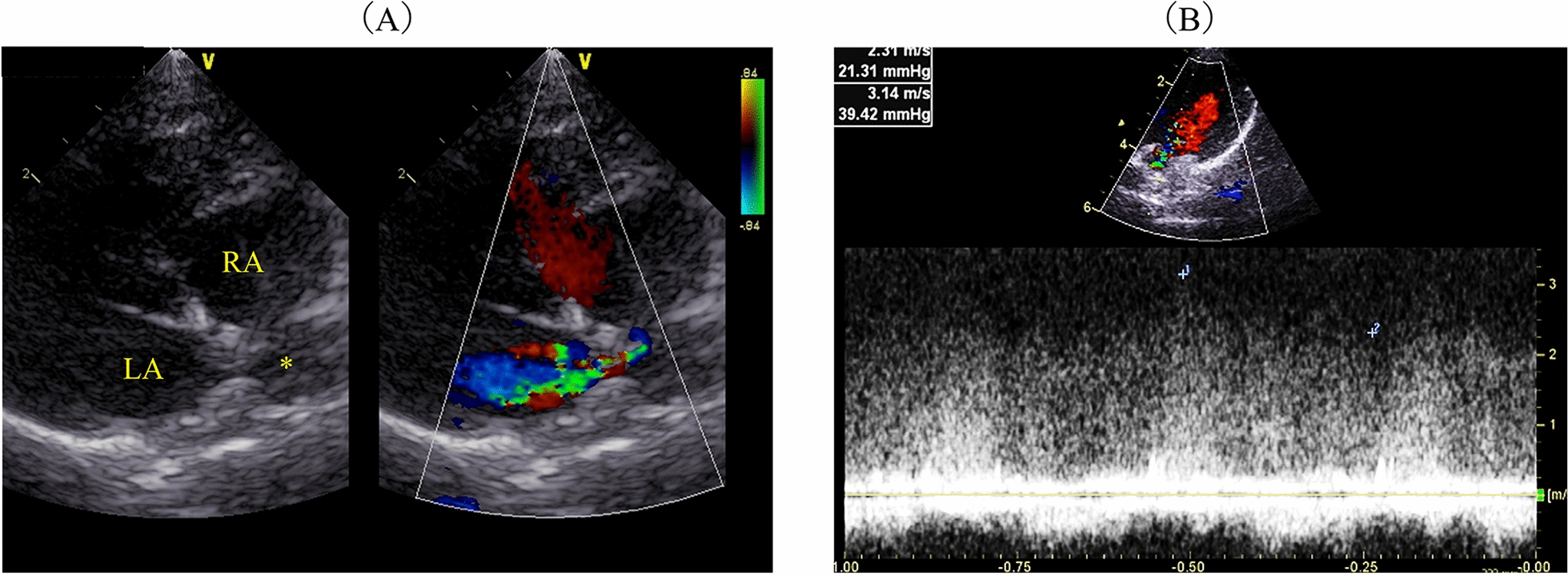
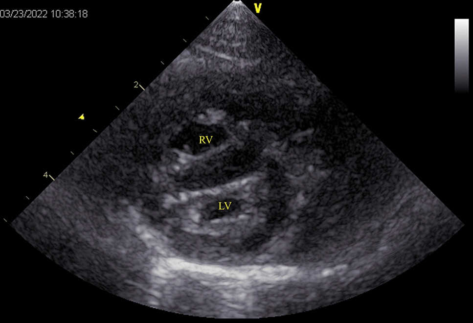
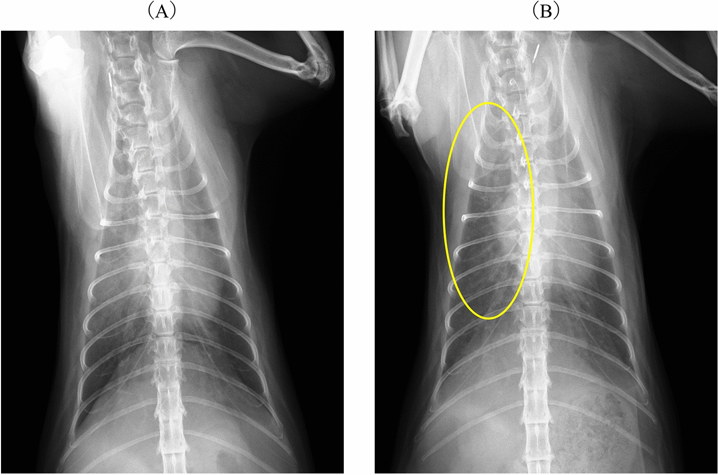
Case presentation: A 2 year-old Norwegian Forest cat was diagnosed with pulmonary oedema and PH secondary to cor triatriatum sinister (CTS) and showed improved breathing following two subcutaneous furosemide treatments, 1 and 2 mg/kg, during an overnight stay at the referral veterinary hospital. Sildenafil alone (0.69 mg/kg, PO, BID) was prescribed post-discharge to address PH without diuretics. Post-discharge from the referral veterinary hospital, collapse and pre-syncope were suspected to be due to PH. Consequently, sildenafil was titrated weekly, starting at 1.09 mg/kg BID and increasing to 1.63 mg/kg BID. Pre-syncope and collapse resolved, and pulmonary opacities reduced considerably, although concerns remained that increased pulmonary blood flow to suspected CTS from sildenafil might worsen cardiogenic pulmonary oedema. The patient was also treated with rivaroxaban (2.5 mg/head, SID), considering the increased risk of thrombus formation due to blood flow stasis and endothelial damage. Thirty-eight days later, the cat presented for the first time to our hosipital (Azabu University Veterinary Teaching Hospital) for examination. On echocardiography, a continuous mosaic blood flow (maximum and minimum velocity, 3.14 m/s; estimated pressure gradient, 39.4 mmHg) was observed in two enlarged pulmonary veins. Pulmonary artery enlargement (main pulmonary artery to thoracic aorta ratio: 1.90), pulmonary vein stenosis (PVS), and diffuse bilateral ground-glass lung opacities were observed using computed tomography. PH with unilateral PVS involving two out of the three right pulmonary veins, specifically the right cranial and right middle pulmonary veins, along with pulmonary parenchymal disease, was diagnosed. The cat was further treated with furosemide (1 mg/kg, BID, PO) with no clinical signs but succumbed to acute dyspnoea 51 days after the first visit.
Conclusions: Unilateral PVS should be considered in young cats with a localised alveolar pattern and no left atrial enlargement, because the prognosis may be poor. Severe PH with PVS may coexist with lung disease. If sildenafil is used, it should be started at a low dose and monitored closely.
期刊介绍:
Acta Veterinaria Scandinavica is an open access journal encompassing all aspects of veterinary research and medicine of domestic and wild animals.

 求助内容:
求助内容: 应助结果提醒方式:


