Ira Komara, Furi Andanawari, Agus Susanto, Euis Reni Yuslianti, Ina Hendiani, Prajna Metta, Amaliya Amaliya
{"title":"胶原-壳聚糖-聚乙烯醇(PVA)膜的生物降解、血管生成和炎症反应:引导组织再生的体内模型","authors":"Ira Komara, Furi Andanawari, Agus Susanto, Euis Reni Yuslianti, Ina Hendiani, Prajna Metta, Amaliya Amaliya","doi":"10.1055/s-0044-1801305","DOIUrl":null,"url":null,"abstract":"<p><p>The aim of this study was to examine the biodegradation, angiogenesis, and inflammatory response in collagen-chitosan-polyvinyl alcohol (PVA) membranes.This study employed an experimental approach utilizing a randomized controlled trial design. Wistar rats were used as subjects, with 51 rats divided into three groups. Each group received a different treatment: application of the collagen-chitosan-PVA membrane, pericardial membrane, or cross-linked pericardial membrane, administered subcutaneously. On days 0, 7, 14, and 30, the rats were terminated, and the membranes and surrounding tissues were collected for analysis. A histological examination was performed to evaluate the membrane biodegradation rate, the number of blood vessels formed, and the inflammatory response.The data were analyzed using the Kruskal-Wallis and Mann-Whitney tests, with a <i>p</i>-value of < 0.05 considered statistically significant.The collagen-chitosan-PVA membrane remained in the tissue up to day 30, while the pericardial membrane and cross-linked pericardial membrane were completely degraded by day 7. The average number of new blood vessels formed in the collagen-chitosan-PVA membrane on days 7, 14, and 30 was greater than that in the pericardial membrane and cross-linked pericardial membrane, which was statistically significant (<i>p</i> < 0.005). The average number of inflammatory cells in the collagen-chitosan-PVA membrane on day 30 was lower than that in the pericardial membrane and cross-linked pericardial membrane, which was statistically significant (<i>p</i> < 0.005) for neutrophils, monocytes, and lymphocytes. However, the difference was not statistically significant (<i>p</i> > 0.05) for eosinophils and mast cells.Biodegradation, angiogenesis, and the inflammatory response in collagen-chitosan-PVA membranes showed better results compared with other membranes. Collagen-chitosan-PVA membranes exhibit potential for application in guided tissue regeneration treatment for periodontal disease.</p>","PeriodicalId":12028,"journal":{"name":"European Journal of Dentistry","volume":" ","pages":"835-842"},"PeriodicalIF":2.1000,"publicationDate":"2025-07-01","publicationTypes":"Journal Article","fieldsOfStudy":null,"isOpenAccess":false,"openAccessPdf":"https://www.ncbi.nlm.nih.gov/pmc/articles/PMC12182392/pdf/","citationCount":"0","resultStr":"{\"title\":\"Biodegradation, Angiogenesis, and Inflammatory Response of a Collagen-Chitosan-Polyvinyl Alcohol (PVA) Membrane: In Vivo Model of Guided Tissue Regeneration.\",\"authors\":\"Ira Komara, Furi Andanawari, Agus Susanto, Euis Reni Yuslianti, Ina Hendiani, Prajna Metta, Amaliya Amaliya\",\"doi\":\"10.1055/s-0044-1801305\",\"DOIUrl\":null,\"url\":null,\"abstract\":\"<p><p>The aim of this study was to examine the biodegradation, angiogenesis, and inflammatory response in collagen-chitosan-polyvinyl alcohol (PVA) membranes.This study employed an experimental approach utilizing a randomized controlled trial design. Wistar rats were used as subjects, with 51 rats divided into three groups. Each group received a different treatment: application of the collagen-chitosan-PVA membrane, pericardial membrane, or cross-linked pericardial membrane, administered subcutaneously. On days 0, 7, 14, and 30, the rats were terminated, and the membranes and surrounding tissues were collected for analysis. A histological examination was performed to evaluate the membrane biodegradation rate, the number of blood vessels formed, and the inflammatory response.The data were analyzed using the Kruskal-Wallis and Mann-Whitney tests, with a <i>p</i>-value of < 0.05 considered statistically significant.The collagen-chitosan-PVA membrane remained in the tissue up to day 30, while the pericardial membrane and cross-linked pericardial membrane were completely degraded by day 7. The average number of new blood vessels formed in the collagen-chitosan-PVA membrane on days 7, 14, and 30 was greater than that in the pericardial membrane and cross-linked pericardial membrane, which was statistically significant (<i>p</i> < 0.005). The average number of inflammatory cells in the collagen-chitosan-PVA membrane on day 30 was lower than that in the pericardial membrane and cross-linked pericardial membrane, which was statistically significant (<i>p</i> < 0.005) for neutrophils, monocytes, and lymphocytes. However, the difference was not statistically significant (<i>p</i> > 0.05) for eosinophils and mast cells.Biodegradation, angiogenesis, and the inflammatory response in collagen-chitosan-PVA membranes showed better results compared with other membranes. Collagen-chitosan-PVA membranes exhibit potential for application in guided tissue regeneration treatment for periodontal disease.</p>\",\"PeriodicalId\":12028,\"journal\":{\"name\":\"European Journal of Dentistry\",\"volume\":\" \",\"pages\":\"835-842\"},\"PeriodicalIF\":2.1000,\"publicationDate\":\"2025-07-01\",\"publicationTypes\":\"Journal Article\",\"fieldsOfStudy\":null,\"isOpenAccess\":false,\"openAccessPdf\":\"https://www.ncbi.nlm.nih.gov/pmc/articles/PMC12182392/pdf/\",\"citationCount\":\"0\",\"resultStr\":null,\"platform\":\"Semanticscholar\",\"paperid\":null,\"PeriodicalName\":\"European Journal of Dentistry\",\"FirstCategoryId\":\"1085\",\"ListUrlMain\":\"https://doi.org/10.1055/s-0044-1801305\",\"RegionNum\":0,\"RegionCategory\":null,\"ArticlePicture\":[],\"TitleCN\":null,\"AbstractTextCN\":null,\"PMCID\":null,\"EPubDate\":\"2025/4/16 0:00:00\",\"PubModel\":\"Epub\",\"JCR\":\"Q1\",\"JCRName\":\"Dentistry\",\"Score\":null,\"Total\":0}","platform":"Semanticscholar","paperid":null,"PeriodicalName":"European Journal of Dentistry","FirstCategoryId":"1085","ListUrlMain":"https://doi.org/10.1055/s-0044-1801305","RegionNum":0,"RegionCategory":null,"ArticlePicture":[],"TitleCN":null,"AbstractTextCN":null,"PMCID":null,"EPubDate":"2025/4/16 0:00:00","PubModel":"Epub","JCR":"Q1","JCRName":"Dentistry","Score":null,"Total":0}
引用次数: 0
摘要
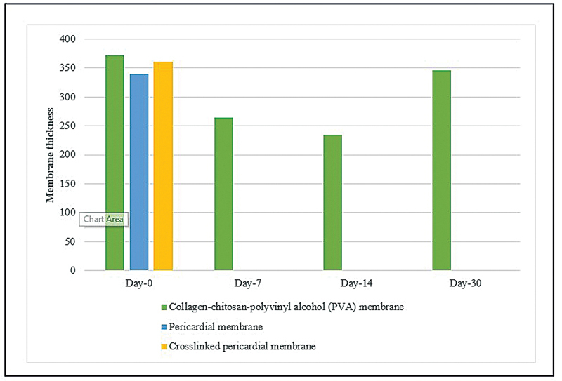
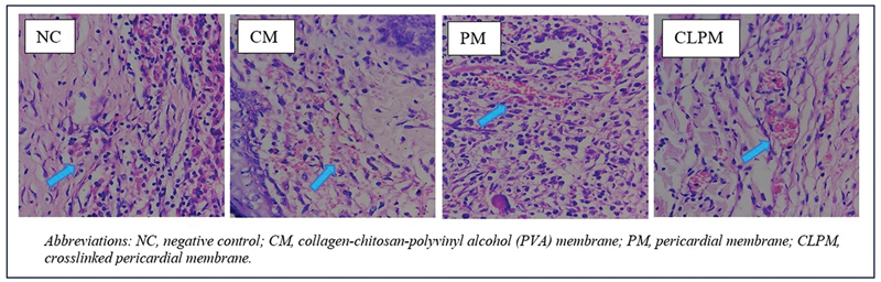
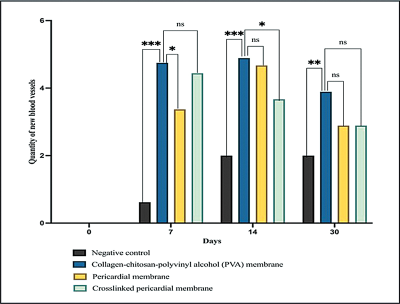
本研究的目的是研究胶原-壳聚糖-聚乙烯醇(PVA)膜的生物降解、血管生成和炎症反应。本研究采用随机对照试验设计的实验方法。以Wistar大鼠为研究对象,将51只大鼠分为三组。各组接受不同的治疗:应用胶原-壳聚糖- pva膜、心包膜或交联心包膜皮下给药。于第0、7、14、30天处死大鼠,收集膜及周围组织进行分析。进行组织学检查以评估膜的生物降解率,形成的血管数量和炎症反应。采用Kruskal-Wallis检验和Mann-Whitney检验对数据进行分析,p值< 0.05认为有统计学意义。胶原-壳聚糖- pva膜在组织中保留至第30天,而心包膜和交联心包膜在第7天完全降解。胶原-壳聚糖- pva膜在第7、14、30天的平均新生血管数均高于心包膜和交联心包膜,嗜酸性粒细胞和肥大细胞的新生血管数差异有统计学意义(p p p > 0.05)。胶原-壳聚糖-聚乙烯醇膜在生物降解、血管生成和炎症反应方面表现出比其他膜更好的效果。胶原-壳聚糖-聚乙烯醇膜在牙周病引导组织再生治疗中具有潜在的应用前景。
Biodegradation, Angiogenesis, and Inflammatory Response of a Collagen-Chitosan-Polyvinyl Alcohol (PVA) Membrane: In Vivo Model of Guided Tissue Regeneration.
The aim of this study was to examine the biodegradation, angiogenesis, and inflammatory response in collagen-chitosan-polyvinyl alcohol (PVA) membranes.This study employed an experimental approach utilizing a randomized controlled trial design. Wistar rats were used as subjects, with 51 rats divided into three groups. Each group received a different treatment: application of the collagen-chitosan-PVA membrane, pericardial membrane, or cross-linked pericardial membrane, administered subcutaneously. On days 0, 7, 14, and 30, the rats were terminated, and the membranes and surrounding tissues were collected for analysis. A histological examination was performed to evaluate the membrane biodegradation rate, the number of blood vessels formed, and the inflammatory response.The data were analyzed using the Kruskal-Wallis and Mann-Whitney tests, with a p-value of < 0.05 considered statistically significant.The collagen-chitosan-PVA membrane remained in the tissue up to day 30, while the pericardial membrane and cross-linked pericardial membrane were completely degraded by day 7. The average number of new blood vessels formed in the collagen-chitosan-PVA membrane on days 7, 14, and 30 was greater than that in the pericardial membrane and cross-linked pericardial membrane, which was statistically significant (p < 0.005). The average number of inflammatory cells in the collagen-chitosan-PVA membrane on day 30 was lower than that in the pericardial membrane and cross-linked pericardial membrane, which was statistically significant (p < 0.005) for neutrophils, monocytes, and lymphocytes. However, the difference was not statistically significant (p > 0.05) for eosinophils and mast cells.Biodegradation, angiogenesis, and the inflammatory response in collagen-chitosan-PVA membranes showed better results compared with other membranes. Collagen-chitosan-PVA membranes exhibit potential for application in guided tissue regeneration treatment for periodontal disease.
期刊介绍:
The European Journal of Dentistry is the official journal of the Dental Investigations Society, based in Turkey. It is a double-blinded peer-reviewed, Open Access, multi-disciplinary international journal addressing various aspects of dentistry. The journal''s board consists of eminent investigators in dentistry from across the globe and presents an ideal international composition. The journal encourages its authors to submit original investigations, reviews, and reports addressing various divisions of dentistry including oral pathology, prosthodontics, endodontics, orthodontics etc. It is available both online and in print.

 求助内容:
求助内容: 应助结果提醒方式:


