{"title":"预测no的机器学习模型的开发与验证。253例左侧结直肠癌淋巴结转移的临床及ct放射学特征分析","authors":"Hongwei Zhang, Kexin Wang, Shurong Liu, Guowei Chen, Yong Jiang, Yingchao Wu, Xiaocong Pang, Xiaoying Wang, Junling Zhang, Xin Wang","doi":"10.1186/s40644-025-00876-y","DOIUrl":null,"url":null,"abstract":"<p><strong>Background: </strong>The appropriate ligation level of the inferior mesenteric artery (IMA) in left-sided colorectal cancer (CRC) surgery is debated, with metastasis in No. 253 lymph node (No. 253 LN) being a key determining factor. This study aimed to develop a machine learning model for predicting metastasis in No. 253 LN.</p><p><strong>Methods: </strong>We retrospectively collected clinical data from 2,118 patients with left-sided CRC and contrast-enhanced CT images from 310 of these patients. From this data, a test set, a training set, and a temporal validation set were constructed. Logistic regression models were used to develop a clinical model, a CT model, and a radiomics model, which were then integrated into a combined model using logical rules. Finally, these models were evaluated using metrics such as the area under the receiver operating characteristic curve (AUC), precision-recall (PR) curves, decision curve analysis (DCA), net reclassification improvement (NRI), and integrated discrimination improvement (IDI).</p><p><strong>Results: </strong>A clinical model, a CT model, and a radiomics model were constructed using univariate logistic regression. A combined model was developed by integrating the clinical, CT, and radiomics models, with positivity defined as all three models being positive at a 90% sensitivity threshold. The clinical model included six predictive factors: tumor site, endoscopic obstruction, CEA levels, growth type, differentiation grade, and pathological classification. The CT model utilized largest lymph node average CT value, short-axis diameter and long-axis diameter. The radiomics model incorporated maximum gray level intensity within the region of interest, large area high gray level emphasis, small area high gray level emphasis and surface area to volume ratio. In the test set, the AUCs for the clinical, CT, radiomics, and combined models were 0.694, 0.663, 0.72, and 0.663, respectively, while in the temporal validation set, they were 0.743, 0.629, 0.716, and 0.8. Specifically, the combined model demonstrated a sensitivity of 0.8 and a specificity of 0.8 in the temporal validation set. By comparing the PR and DCA curves, the combined model demonstrated better performance. Additionally, the combined model showed moderate improvements in INR and IDI compared to other models.</p><p><strong>Conclusion: </strong>A clinical and CT-based radiomics model shows promise in predicting No. 253 LN metastasis in left-sided CRC and provides insights for optimizing IMA ligation strategies.</p>","PeriodicalId":9548,"journal":{"name":"Cancer Imaging","volume":"25 1","pages":"57"},"PeriodicalIF":3.5000,"publicationDate":"2025-04-29","publicationTypes":"Journal Article","fieldsOfStudy":null,"isOpenAccess":false,"openAccessPdf":"https://www.ncbi.nlm.nih.gov/pmc/articles/PMC12039209/pdf/","citationCount":"0","resultStr":"{\"title\":\"Development and validation of machine learning models for predicting no. 253 lymph node metastasis in left-sided colorectal cancer using clinical and CT-based radiomic features.\",\"authors\":\"Hongwei Zhang, Kexin Wang, Shurong Liu, Guowei Chen, Yong Jiang, Yingchao Wu, Xiaocong Pang, Xiaoying Wang, Junling Zhang, Xin Wang\",\"doi\":\"10.1186/s40644-025-00876-y\",\"DOIUrl\":null,\"url\":null,\"abstract\":\"<p><strong>Background: </strong>The appropriate ligation level of the inferior mesenteric artery (IMA) in left-sided colorectal cancer (CRC) surgery is debated, with metastasis in No. 253 lymph node (No. 253 LN) being a key determining factor. This study aimed to develop a machine learning model for predicting metastasis in No. 253 LN.</p><p><strong>Methods: </strong>We retrospectively collected clinical data from 2,118 patients with left-sided CRC and contrast-enhanced CT images from 310 of these patients. From this data, a test set, a training set, and a temporal validation set were constructed. Logistic regression models were used to develop a clinical model, a CT model, and a radiomics model, which were then integrated into a combined model using logical rules. Finally, these models were evaluated using metrics such as the area under the receiver operating characteristic curve (AUC), precision-recall (PR) curves, decision curve analysis (DCA), net reclassification improvement (NRI), and integrated discrimination improvement (IDI).</p><p><strong>Results: </strong>A clinical model, a CT model, and a radiomics model were constructed using univariate logistic regression. A combined model was developed by integrating the clinical, CT, and radiomics models, with positivity defined as all three models being positive at a 90% sensitivity threshold. The clinical model included six predictive factors: tumor site, endoscopic obstruction, CEA levels, growth type, differentiation grade, and pathological classification. The CT model utilized largest lymph node average CT value, short-axis diameter and long-axis diameter. The radiomics model incorporated maximum gray level intensity within the region of interest, large area high gray level emphasis, small area high gray level emphasis and surface area to volume ratio. In the test set, the AUCs for the clinical, CT, radiomics, and combined models were 0.694, 0.663, 0.72, and 0.663, respectively, while in the temporal validation set, they were 0.743, 0.629, 0.716, and 0.8. Specifically, the combined model demonstrated a sensitivity of 0.8 and a specificity of 0.8 in the temporal validation set. By comparing the PR and DCA curves, the combined model demonstrated better performance. Additionally, the combined model showed moderate improvements in INR and IDI compared to other models.</p><p><strong>Conclusion: </strong>A clinical and CT-based radiomics model shows promise in predicting No. 253 LN metastasis in left-sided CRC and provides insights for optimizing IMA ligation strategies.</p>\",\"PeriodicalId\":9548,\"journal\":{\"name\":\"Cancer Imaging\",\"volume\":\"25 1\",\"pages\":\"57\"},\"PeriodicalIF\":3.5000,\"publicationDate\":\"2025-04-29\",\"publicationTypes\":\"Journal Article\",\"fieldsOfStudy\":null,\"isOpenAccess\":false,\"openAccessPdf\":\"https://www.ncbi.nlm.nih.gov/pmc/articles/PMC12039209/pdf/\",\"citationCount\":\"0\",\"resultStr\":null,\"platform\":\"Semanticscholar\",\"paperid\":null,\"PeriodicalName\":\"Cancer Imaging\",\"FirstCategoryId\":\"3\",\"ListUrlMain\":\"https://doi.org/10.1186/s40644-025-00876-y\",\"RegionNum\":2,\"RegionCategory\":\"医学\",\"ArticlePicture\":[],\"TitleCN\":null,\"AbstractTextCN\":null,\"PMCID\":null,\"EPubDate\":\"\",\"PubModel\":\"\",\"JCR\":\"Q2\",\"JCRName\":\"ONCOLOGY\",\"Score\":null,\"Total\":0}","platform":"Semanticscholar","paperid":null,"PeriodicalName":"Cancer Imaging","FirstCategoryId":"3","ListUrlMain":"https://doi.org/10.1186/s40644-025-00876-y","RegionNum":2,"RegionCategory":"医学","ArticlePicture":[],"TitleCN":null,"AbstractTextCN":null,"PMCID":null,"EPubDate":"","PubModel":"","JCR":"Q2","JCRName":"ONCOLOGY","Score":null,"Total":0}
Development and validation of machine learning models for predicting no. 253 lymph node metastasis in left-sided colorectal cancer using clinical and CT-based radiomic features.
Background: The appropriate ligation level of the inferior mesenteric artery (IMA) in left-sided colorectal cancer (CRC) surgery is debated, with metastasis in No. 253 lymph node (No. 253 LN) being a key determining factor. This study aimed to develop a machine learning model for predicting metastasis in No. 253 LN.
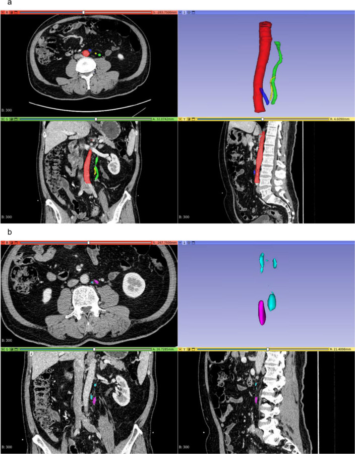
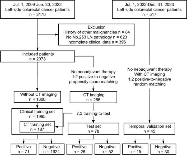
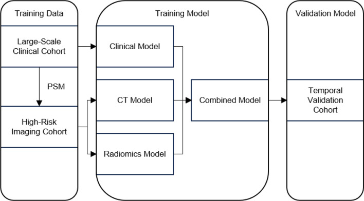
Methods: We retrospectively collected clinical data from 2,118 patients with left-sided CRC and contrast-enhanced CT images from 310 of these patients. From this data, a test set, a training set, and a temporal validation set were constructed. Logistic regression models were used to develop a clinical model, a CT model, and a radiomics model, which were then integrated into a combined model using logical rules. Finally, these models were evaluated using metrics such as the area under the receiver operating characteristic curve (AUC), precision-recall (PR) curves, decision curve analysis (DCA), net reclassification improvement (NRI), and integrated discrimination improvement (IDI).
Results: A clinical model, a CT model, and a radiomics model were constructed using univariate logistic regression. A combined model was developed by integrating the clinical, CT, and radiomics models, with positivity defined as all three models being positive at a 90% sensitivity threshold. The clinical model included six predictive factors: tumor site, endoscopic obstruction, CEA levels, growth type, differentiation grade, and pathological classification. The CT model utilized largest lymph node average CT value, short-axis diameter and long-axis diameter. The radiomics model incorporated maximum gray level intensity within the region of interest, large area high gray level emphasis, small area high gray level emphasis and surface area to volume ratio. In the test set, the AUCs for the clinical, CT, radiomics, and combined models were 0.694, 0.663, 0.72, and 0.663, respectively, while in the temporal validation set, they were 0.743, 0.629, 0.716, and 0.8. Specifically, the combined model demonstrated a sensitivity of 0.8 and a specificity of 0.8 in the temporal validation set. By comparing the PR and DCA curves, the combined model demonstrated better performance. Additionally, the combined model showed moderate improvements in INR and IDI compared to other models.
Conclusion: A clinical and CT-based radiomics model shows promise in predicting No. 253 LN metastasis in left-sided CRC and provides insights for optimizing IMA ligation strategies.
Cancer ImagingONCOLOGY-RADIOLOGY, NUCLEAR MEDICINE & MEDICAL IMAGING
CiteScore
7.00
自引率
0.00%
发文量
66
审稿时长
>12 weeks
期刊介绍:
Cancer Imaging is an open access, peer-reviewed journal publishing original articles, reviews and editorials written by expert international radiologists working in oncology.
The journal encompasses CT, MR, PET, ultrasound, radionuclide and multimodal imaging in all kinds of malignant tumours, plus new developments, techniques and innovations. Topics of interest include:
Breast Imaging
Chest
Complications of treatment
Ear, Nose & Throat
Gastrointestinal
Hepatobiliary & Pancreatic
Imaging biomarkers
Interventional
Lymphoma
Measurement of tumour response
Molecular functional imaging
Musculoskeletal
Neuro oncology
Nuclear Medicine
Paediatric.

 求助内容:
求助内容: 应助结果提醒方式:


