{"title":"脉络膜丛体积与多发性硬化症的相关性。","authors":"Britta Krieger, Barbara Bellenberg, Anna Katharina Roenneke, Ruth Schneider, Theodoros Ladopoulos, Zainab Abbas, Rebekka Rust, Tanja Schmitz-Hübsch, Claudia Chien, Ralf Gold, Friedemann Paul, Carsten Lukas","doi":"10.1186/s12987-025-00656-7","DOIUrl":null,"url":null,"abstract":"<p><strong>Background: </strong>The choroid plexus (ChP) plays a pivotal role in inflammatory processes that occur in multiple sclerosis (MS). The enlargement of the ChP in relapsing-remitting multiple sclerosis (RRMS) is considered to be an indication of disease activity and has been associated with periventricular remyelination failure. This cross-sectional study aimed to identify the relationship between ChP and periventricular tissue damage which occurs in MS, and to elucidate the role of neuroinflammation in primary progressive multiple sclerosis (PPMS).</p><p><strong>Methods: </strong>ChP volume was assessed by a novel deep learning segmentation method based on structural MRI data acquired from two centers. In total, 141 RRMS and 64 PPMS patients were included, along with 75 healthy control subjects. In addition, T1w/FLAIR ratios were calculated within periventricular bands to quantify microstructural tissue damage and to assess its relationship to ChP volume.</p><p><strong>Results: </strong>When compared to healthy controls, ChP volumes were significantly increased in RRMS, but not in patients with PPMS. T1w/FLAIR ratios in the normal appearing white matter (NAWM) showing periventricular gradients were decreased in patients with multiple sclerosis when compared to healthy control subjects and lower T1w/FLAIR ratios radiating out from the lateral ventricles were found in patients with PPMS. A relationship between ChP volume and T1w/FLAIR ratio in NAWM was found within the inner periventricular bands in RRMS patients. A longer duration of disease was associated with larger ChP volumes only in RRMS patients. Enlarged ChP volumes were also significantly associated with reduced cortex volumes and increased lesion volumes in RRMS.</p><p><strong>Conclusions: </strong>Our analysis confirmed that the ChP was significantly enlarged in patients with RRMS, which was related to brain lesion volumes and which suggested a dynamic development as it was associated with disease duration. Plexus enlargement was further associated with periventricular demyelination or tissue damage assessed by T1w/FLAIR ratios in RRMS. Furthermore, we did not find an enlargement of the ChP in patients with PPMS, possibly indicating the reduced involvement of inflammatory processes in the progressive phase of MS. The association between enlarged ChP volumes and cortical atrophy in RRMS highlighted the vulnerability of structures close to the CSF.</p>","PeriodicalId":12321,"journal":{"name":"Fluids and Barriers of the CNS","volume":"22 1","pages":"47"},"PeriodicalIF":6.2000,"publicationDate":"2025-05-08","publicationTypes":"Journal Article","fieldsOfStudy":null,"isOpenAccess":false,"openAccessPdf":"https://www.ncbi.nlm.nih.gov/pmc/articles/PMC12060557/pdf/","citationCount":"0","resultStr":"{\"title\":\"Relevance of choroid plexus volumes in multiple sclerosis.\",\"authors\":\"Britta Krieger, Barbara Bellenberg, Anna Katharina Roenneke, Ruth Schneider, Theodoros Ladopoulos, Zainab Abbas, Rebekka Rust, Tanja Schmitz-Hübsch, Claudia Chien, Ralf Gold, Friedemann Paul, Carsten Lukas\",\"doi\":\"10.1186/s12987-025-00656-7\",\"DOIUrl\":null,\"url\":null,\"abstract\":\"<p><strong>Background: </strong>The choroid plexus (ChP) plays a pivotal role in inflammatory processes that occur in multiple sclerosis (MS). The enlargement of the ChP in relapsing-remitting multiple sclerosis (RRMS) is considered to be an indication of disease activity and has been associated with periventricular remyelination failure. This cross-sectional study aimed to identify the relationship between ChP and periventricular tissue damage which occurs in MS, and to elucidate the role of neuroinflammation in primary progressive multiple sclerosis (PPMS).</p><p><strong>Methods: </strong>ChP volume was assessed by a novel deep learning segmentation method based on structural MRI data acquired from two centers. In total, 141 RRMS and 64 PPMS patients were included, along with 75 healthy control subjects. In addition, T1w/FLAIR ratios were calculated within periventricular bands to quantify microstructural tissue damage and to assess its relationship to ChP volume.</p><p><strong>Results: </strong>When compared to healthy controls, ChP volumes were significantly increased in RRMS, but not in patients with PPMS. T1w/FLAIR ratios in the normal appearing white matter (NAWM) showing periventricular gradients were decreased in patients with multiple sclerosis when compared to healthy control subjects and lower T1w/FLAIR ratios radiating out from the lateral ventricles were found in patients with PPMS. A relationship between ChP volume and T1w/FLAIR ratio in NAWM was found within the inner periventricular bands in RRMS patients. A longer duration of disease was associated with larger ChP volumes only in RRMS patients. Enlarged ChP volumes were also significantly associated with reduced cortex volumes and increased lesion volumes in RRMS.</p><p><strong>Conclusions: </strong>Our analysis confirmed that the ChP was significantly enlarged in patients with RRMS, which was related to brain lesion volumes and which suggested a dynamic development as it was associated with disease duration. Plexus enlargement was further associated with periventricular demyelination or tissue damage assessed by T1w/FLAIR ratios in RRMS. Furthermore, we did not find an enlargement of the ChP in patients with PPMS, possibly indicating the reduced involvement of inflammatory processes in the progressive phase of MS. The association between enlarged ChP volumes and cortical atrophy in RRMS highlighted the vulnerability of structures close to the CSF.</p>\",\"PeriodicalId\":12321,\"journal\":{\"name\":\"Fluids and Barriers of the CNS\",\"volume\":\"22 1\",\"pages\":\"47\"},\"PeriodicalIF\":6.2000,\"publicationDate\":\"2025-05-08\",\"publicationTypes\":\"Journal Article\",\"fieldsOfStudy\":null,\"isOpenAccess\":false,\"openAccessPdf\":\"https://www.ncbi.nlm.nih.gov/pmc/articles/PMC12060557/pdf/\",\"citationCount\":\"0\",\"resultStr\":null,\"platform\":\"Semanticscholar\",\"paperid\":null,\"PeriodicalName\":\"Fluids and Barriers of the CNS\",\"FirstCategoryId\":\"3\",\"ListUrlMain\":\"https://doi.org/10.1186/s12987-025-00656-7\",\"RegionNum\":1,\"RegionCategory\":\"医学\",\"ArticlePicture\":[],\"TitleCN\":null,\"AbstractTextCN\":null,\"PMCID\":null,\"EPubDate\":\"\",\"PubModel\":\"\",\"JCR\":\"Q1\",\"JCRName\":\"NEUROSCIENCES\",\"Score\":null,\"Total\":0}","platform":"Semanticscholar","paperid":null,"PeriodicalName":"Fluids and Barriers of the CNS","FirstCategoryId":"3","ListUrlMain":"https://doi.org/10.1186/s12987-025-00656-7","RegionNum":1,"RegionCategory":"医学","ArticlePicture":[],"TitleCN":null,"AbstractTextCN":null,"PMCID":null,"EPubDate":"","PubModel":"","JCR":"Q1","JCRName":"NEUROSCIENCES","Score":null,"Total":0}
Relevance of choroid plexus volumes in multiple sclerosis.
Background: The choroid plexus (ChP) plays a pivotal role in inflammatory processes that occur in multiple sclerosis (MS). The enlargement of the ChP in relapsing-remitting multiple sclerosis (RRMS) is considered to be an indication of disease activity and has been associated with periventricular remyelination failure. This cross-sectional study aimed to identify the relationship between ChP and periventricular tissue damage which occurs in MS, and to elucidate the role of neuroinflammation in primary progressive multiple sclerosis (PPMS).
Methods: ChP volume was assessed by a novel deep learning segmentation method based on structural MRI data acquired from two centers. In total, 141 RRMS and 64 PPMS patients were included, along with 75 healthy control subjects. In addition, T1w/FLAIR ratios were calculated within periventricular bands to quantify microstructural tissue damage and to assess its relationship to ChP volume.
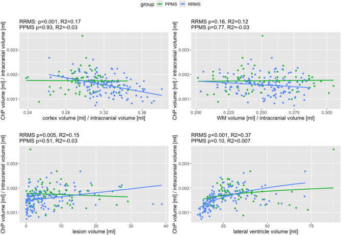
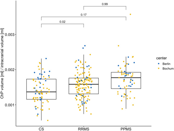
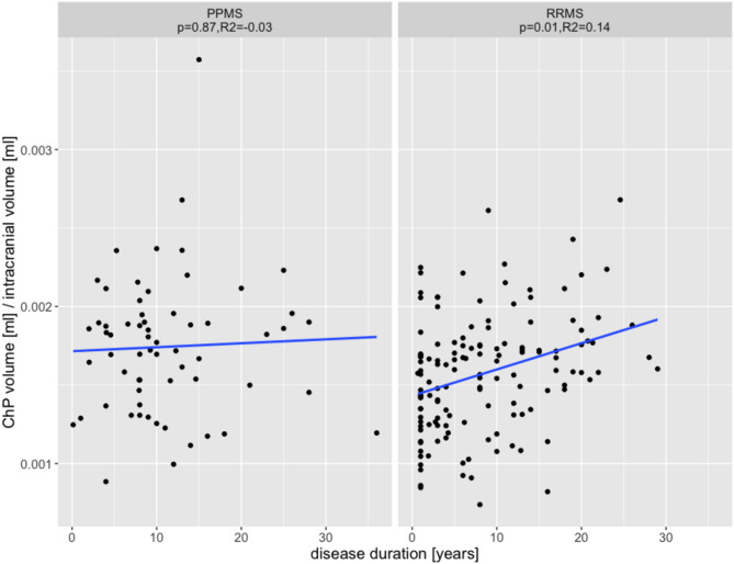
Results: When compared to healthy controls, ChP volumes were significantly increased in RRMS, but not in patients with PPMS. T1w/FLAIR ratios in the normal appearing white matter (NAWM) showing periventricular gradients were decreased in patients with multiple sclerosis when compared to healthy control subjects and lower T1w/FLAIR ratios radiating out from the lateral ventricles were found in patients with PPMS. A relationship between ChP volume and T1w/FLAIR ratio in NAWM was found within the inner periventricular bands in RRMS patients. A longer duration of disease was associated with larger ChP volumes only in RRMS patients. Enlarged ChP volumes were also significantly associated with reduced cortex volumes and increased lesion volumes in RRMS.
Conclusions: Our analysis confirmed that the ChP was significantly enlarged in patients with RRMS, which was related to brain lesion volumes and which suggested a dynamic development as it was associated with disease duration. Plexus enlargement was further associated with periventricular demyelination or tissue damage assessed by T1w/FLAIR ratios in RRMS. Furthermore, we did not find an enlargement of the ChP in patients with PPMS, possibly indicating the reduced involvement of inflammatory processes in the progressive phase of MS. The association between enlarged ChP volumes and cortical atrophy in RRMS highlighted the vulnerability of structures close to the CSF.
期刊介绍:
"Fluids and Barriers of the CNS" is a scholarly open access journal that specializes in the intricate world of the central nervous system's fluids and barriers, which are pivotal for the health and well-being of the human body. This journal is a peer-reviewed platform that welcomes research manuscripts exploring the full spectrum of CNS fluids and barriers, with a particular focus on their roles in both health and disease.
At the heart of this journal's interest is the cerebrospinal fluid (CSF), a vital fluid that circulates within the brain and spinal cord, playing a multifaceted role in the normal functioning of the brain and in various neurological conditions. The journal delves into the composition, circulation, and absorption of CSF, as well as its relationship with the parenchymal interstitial fluid and the neurovascular unit at the blood-brain barrier (BBB).
 求助内容:
求助内容: 应助结果提醒方式:


