{"title":"治疗前与基线相比:在病变摄取和可检出性方面有差异吗?","authors":"Masatoshi Hotta, Daisuke Horikawa, Keiichi Kurihara, Shuhei Ohashi, Kaori Saito, Fuyuki Inagaki","doi":"10.1007/s12149-025-02050-7","DOIUrl":null,"url":null,"abstract":"<div><h3>Background</h3><p><sup>111</sup>In-pentetreotide imaging remains used for pre-treatment screening in peptide receptor radionuclide therapy (PRRT) in regions where somatostatin receptor (SSTR)-PET tracers are clinically unavailable. Post-treatment <sup>177</sup>Lu-DOTATATE imaging at the first PRRT cycle serves as a baseline for response assessment on subsequent post-treatment imaging, paralleling the role of pre-treatment <sup>111</sup>In-pentetreotide imaging. However, differences between these SSTR scans have not been reported. This study aims to compare <sup>111</sup>In-pentetreotide and <sup>177</sup>Lu-DOTATATE imaging, focusing on lesion uptake and numbers.</p><h3>Methods</h3><p>This retrospective, single-center study included patients with neuroendocrine tumors (NETs) who underwent PRRT at our hospital. Planar and SPECT/CT of pre-treatment <sup>111</sup>In-pentetreotide and post-treatment <sup>177</sup>Lu-DOTATATE imaging at the first PRRT cycle were analyzed. For visual analysis, the number of SSTR-positive (Krenning score ≥ 2) lesions was counted for each tracer. For quantitative analysis, up to three representative SSTR-positive lesions per patient were selected, and the maximum standardized uptake value (SUVmax) and tumor-to-background (T/B) ratio were measured. Wilcoxon signed-rank test was used to compare lesion counts and uptake parameters.</p><h3>Results</h3><p>A total of 10 patients and 28 lesions were included. The median interval between <sup>111</sup>In-pentetreotide and <sup>177</sup>Lu-DOTATATE imaging was 43 days. <sup>177</sup>Lu-DOTATATE imaging detected significantly more lesions than <sup>111</sup>In-pentetreotide on both planar (median 10 vs. 6.5 lesions, <i>p</i> = 0.009) and SPECT/CT (median 13.5 vs. 8 lesions, <i>p</i> = 0.014). Lesion uptake was higher with <sup>177</sup>Lu-DOTATATE, with SUVmax (median 4.5 vs. 2.9, <i>p</i> < 0.001) and T/B ratios (median 13.9 vs. 4.7, <i>p</i> < 0.001). Sub-centimeter lesions accounted for most of the additional detections on <sup>177</sup>Lu-DOTATATE SPECT/CT.</p><h3>Conclusion</h3><p>Baseline <sup>177</sup>Lu-DOTATATE imaging demonstrated higher lesion uptake and detected more lesions compared to pre-treatment <sup>111</sup>In-pentetreotide imaging. Careful interpretation of baseline <sup>177</sup>Lu-DOTATATE imaging is essential to avoid misinterpreting the increased number of detected lesions as disease progression. Screening with <sup>111</sup>In-pentetreotide imaging may underestimate treatable lesions by PRRT, particularly when planar imaging alone is used, highlighting the need for SSTR PET in pre-treatment evaluation.</p></div>","PeriodicalId":8007,"journal":{"name":"Annals of Nuclear Medicine","volume":"39 7","pages":"747 - 753"},"PeriodicalIF":2.5000,"publicationDate":"2025-04-14","publicationTypes":"Journal Article","fieldsOfStudy":null,"isOpenAccess":false,"openAccessPdf":"","citationCount":"0","resultStr":"{\"title\":\"Pre-treatment 111In-pentetreotide SPECT/CT vs. baseline 177Lu-DOTATATE SPECT/CT: are there differences in lesion uptake and detectability?\",\"authors\":\"Masatoshi Hotta, Daisuke Horikawa, Keiichi Kurihara, Shuhei Ohashi, Kaori Saito, Fuyuki Inagaki\",\"doi\":\"10.1007/s12149-025-02050-7\",\"DOIUrl\":null,\"url\":null,\"abstract\":\"<div><h3>Background</h3><p><sup>111</sup>In-pentetreotide imaging remains used for pre-treatment screening in peptide receptor radionuclide therapy (PRRT) in regions where somatostatin receptor (SSTR)-PET tracers are clinically unavailable. Post-treatment <sup>177</sup>Lu-DOTATATE imaging at the first PRRT cycle serves as a baseline for response assessment on subsequent post-treatment imaging, paralleling the role of pre-treatment <sup>111</sup>In-pentetreotide imaging. However, differences between these SSTR scans have not been reported. This study aims to compare <sup>111</sup>In-pentetreotide and <sup>177</sup>Lu-DOTATATE imaging, focusing on lesion uptake and numbers.</p><h3>Methods</h3><p>This retrospective, single-center study included patients with neuroendocrine tumors (NETs) who underwent PRRT at our hospital. Planar and SPECT/CT of pre-treatment <sup>111</sup>In-pentetreotide and post-treatment <sup>177</sup>Lu-DOTATATE imaging at the first PRRT cycle were analyzed. For visual analysis, the number of SSTR-positive (Krenning score ≥ 2) lesions was counted for each tracer. For quantitative analysis, up to three representative SSTR-positive lesions per patient were selected, and the maximum standardized uptake value (SUVmax) and tumor-to-background (T/B) ratio were measured. Wilcoxon signed-rank test was used to compare lesion counts and uptake parameters.</p><h3>Results</h3><p>A total of 10 patients and 28 lesions were included. The median interval between <sup>111</sup>In-pentetreotide and <sup>177</sup>Lu-DOTATATE imaging was 43 days. <sup>177</sup>Lu-DOTATATE imaging detected significantly more lesions than <sup>111</sup>In-pentetreotide on both planar (median 10 vs. 6.5 lesions, <i>p</i> = 0.009) and SPECT/CT (median 13.5 vs. 8 lesions, <i>p</i> = 0.014). Lesion uptake was higher with <sup>177</sup>Lu-DOTATATE, with SUVmax (median 4.5 vs. 2.9, <i>p</i> < 0.001) and T/B ratios (median 13.9 vs. 4.7, <i>p</i> < 0.001). Sub-centimeter lesions accounted for most of the additional detections on <sup>177</sup>Lu-DOTATATE SPECT/CT.</p><h3>Conclusion</h3><p>Baseline <sup>177</sup>Lu-DOTATATE imaging demonstrated higher lesion uptake and detected more lesions compared to pre-treatment <sup>111</sup>In-pentetreotide imaging. Careful interpretation of baseline <sup>177</sup>Lu-DOTATATE imaging is essential to avoid misinterpreting the increased number of detected lesions as disease progression. Screening with <sup>111</sup>In-pentetreotide imaging may underestimate treatable lesions by PRRT, particularly when planar imaging alone is used, highlighting the need for SSTR PET in pre-treatment evaluation.</p></div>\",\"PeriodicalId\":8007,\"journal\":{\"name\":\"Annals of Nuclear Medicine\",\"volume\":\"39 7\",\"pages\":\"747 - 753\"},\"PeriodicalIF\":2.5000,\"publicationDate\":\"2025-04-14\",\"publicationTypes\":\"Journal Article\",\"fieldsOfStudy\":null,\"isOpenAccess\":false,\"openAccessPdf\":\"\",\"citationCount\":\"0\",\"resultStr\":null,\"platform\":\"Semanticscholar\",\"paperid\":null,\"PeriodicalName\":\"Annals of Nuclear Medicine\",\"FirstCategoryId\":\"3\",\"ListUrlMain\":\"https://link.springer.com/article/10.1007/s12149-025-02050-7\",\"RegionNum\":4,\"RegionCategory\":\"医学\",\"ArticlePicture\":[],\"TitleCN\":null,\"AbstractTextCN\":null,\"PMCID\":null,\"EPubDate\":\"\",\"PubModel\":\"\",\"JCR\":\"Q2\",\"JCRName\":\"RADIOLOGY, NUCLEAR MEDICINE & MEDICAL IMAGING\",\"Score\":null,\"Total\":0}","platform":"Semanticscholar","paperid":null,"PeriodicalName":"Annals of Nuclear Medicine","FirstCategoryId":"3","ListUrlMain":"https://link.springer.com/article/10.1007/s12149-025-02050-7","RegionNum":4,"RegionCategory":"医学","ArticlePicture":[],"TitleCN":null,"AbstractTextCN":null,"PMCID":null,"EPubDate":"","PubModel":"","JCR":"Q2","JCRName":"RADIOLOGY, NUCLEAR MEDICINE & MEDICAL IMAGING","Score":null,"Total":0}
Pre-treatment 111In-pentetreotide SPECT/CT vs. baseline 177Lu-DOTATATE SPECT/CT: are there differences in lesion uptake and detectability?
Background
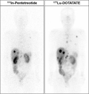
111In-pentetreotide imaging remains used for pre-treatment screening in peptide receptor radionuclide therapy (PRRT) in regions where somatostatin receptor (SSTR)-PET tracers are clinically unavailable. Post-treatment 177Lu-DOTATATE imaging at the first PRRT cycle serves as a baseline for response assessment on subsequent post-treatment imaging, paralleling the role of pre-treatment 111In-pentetreotide imaging. However, differences between these SSTR scans have not been reported. This study aims to compare 111In-pentetreotide and 177Lu-DOTATATE imaging, focusing on lesion uptake and numbers.
Methods
This retrospective, single-center study included patients with neuroendocrine tumors (NETs) who underwent PRRT at our hospital. Planar and SPECT/CT of pre-treatment 111In-pentetreotide and post-treatment 177Lu-DOTATATE imaging at the first PRRT cycle were analyzed. For visual analysis, the number of SSTR-positive (Krenning score ≥ 2) lesions was counted for each tracer. For quantitative analysis, up to three representative SSTR-positive lesions per patient were selected, and the maximum standardized uptake value (SUVmax) and tumor-to-background (T/B) ratio were measured. Wilcoxon signed-rank test was used to compare lesion counts and uptake parameters.
Results
A total of 10 patients and 28 lesions were included. The median interval between 111In-pentetreotide and 177Lu-DOTATATE imaging was 43 days. 177Lu-DOTATATE imaging detected significantly more lesions than 111In-pentetreotide on both planar (median 10 vs. 6.5 lesions, p = 0.009) and SPECT/CT (median 13.5 vs. 8 lesions, p = 0.014). Lesion uptake was higher with 177Lu-DOTATATE, with SUVmax (median 4.5 vs. 2.9, p < 0.001) and T/B ratios (median 13.9 vs. 4.7, p < 0.001). Sub-centimeter lesions accounted for most of the additional detections on 177Lu-DOTATATE SPECT/CT.
Conclusion
Baseline 177Lu-DOTATATE imaging demonstrated higher lesion uptake and detected more lesions compared to pre-treatment 111In-pentetreotide imaging. Careful interpretation of baseline 177Lu-DOTATATE imaging is essential to avoid misinterpreting the increased number of detected lesions as disease progression. Screening with 111In-pentetreotide imaging may underestimate treatable lesions by PRRT, particularly when planar imaging alone is used, highlighting the need for SSTR PET in pre-treatment evaluation.
期刊介绍:
Annals of Nuclear Medicine is an official journal of the Japanese Society of Nuclear Medicine. It develops the appropriate application of radioactive substances and stable nuclides in the field of medicine.
The journal promotes the exchange of ideas and information and research in nuclear medicine and includes the medical application of radionuclides and related subjects. It presents original articles, short communications, reviews and letters to the editor.

 求助内容:
求助内容: 应助结果提醒方式:


