{"title":"使用18f -氟替他莫进行淀粉样蛋白PET检查的脑放射性药物积累的时间变化。","authors":"Shota Takemoto, Koji Onuki, Keiko Tanimoto, Masaho Taniguchi, Takako Suero, Mio Okamoto, Satoshi Kimura, Monami Osawa, Haruka Takeshige-Amano, Noriko Nishikawa, Shigeki Aoki, Ryohei Kuwatsuru, Nobutaka Hattori, Koji Murakami","doi":"10.1007/s12149-025-02046-3","DOIUrl":null,"url":null,"abstract":"<div><h3>Objective</h3><p>The purpose of this study was to examine how the radiopharmaceutical accumulation in the brain changes with the elapsed time between the administration of <sup>18</sup>F-flutemetamol and the start of imaging, and to determine its effect on quantitative indices.</p><h3>Methods</h3><p>The study population consisted of 25 subjects who agreed to participate in the study. After visual evaluation by the radiologist, 14 subjects tested negative for Aβ accumulation, and 11 subjects tested positive as well. The study population was treated with <sup>18</sup>F-flutemeamol, and list mode acquisition was performed for 50 min starting at 60 min after the time of administration. From the acquired list data, five PET images were extracted at 10-min intervals from the start to the end of acquisition, a PET image corresponding to 20 min of acquisition from 60 min after administration, and a PET image corresponding to 20 min of acquisition from 90 min after administration, respectively. Pixel values were measured for the PET images created and quantitative indices (pixel value, SUVr, Centiloid scale) were calculated and compared.</p><h3>Results</h3><p>In most of the PET images, pixel values showed a decreasing trend with elapsed time after radiopharmaceutical administration. Accordingly, calculated SUVr and Centiloid Scale also changed.</p><h3>Conclusions</h3><p>Elapsed time after radiopharmaceutical administration resulted in a washout of the radiopharmaceutical accumulation in the brain. From this, it was suggested that the quantitative indices change.</p></div>","PeriodicalId":8007,"journal":{"name":"Annals of Nuclear Medicine","volume":"39 7","pages":"732 - 746"},"PeriodicalIF":2.5000,"publicationDate":"2025-04-19","publicationTypes":"Journal Article","fieldsOfStudy":null,"isOpenAccess":false,"openAccessPdf":"https://www.ncbi.nlm.nih.gov/pmc/articles/PMC12174224/pdf/","citationCount":"0","resultStr":"{\"title\":\"Elapsed time changes of the brain radiopharmaceutical accumulation of the amyloid PET examination using 18F-flutemetamol\",\"authors\":\"Shota Takemoto, Koji Onuki, Keiko Tanimoto, Masaho Taniguchi, Takako Suero, Mio Okamoto, Satoshi Kimura, Monami Osawa, Haruka Takeshige-Amano, Noriko Nishikawa, Shigeki Aoki, Ryohei Kuwatsuru, Nobutaka Hattori, Koji Murakami\",\"doi\":\"10.1007/s12149-025-02046-3\",\"DOIUrl\":null,\"url\":null,\"abstract\":\"<div><h3>Objective</h3><p>The purpose of this study was to examine how the radiopharmaceutical accumulation in the brain changes with the elapsed time between the administration of <sup>18</sup>F-flutemetamol and the start of imaging, and to determine its effect on quantitative indices.</p><h3>Methods</h3><p>The study population consisted of 25 subjects who agreed to participate in the study. After visual evaluation by the radiologist, 14 subjects tested negative for Aβ accumulation, and 11 subjects tested positive as well. The study population was treated with <sup>18</sup>F-flutemeamol, and list mode acquisition was performed for 50 min starting at 60 min after the time of administration. From the acquired list data, five PET images were extracted at 10-min intervals from the start to the end of acquisition, a PET image corresponding to 20 min of acquisition from 60 min after administration, and a PET image corresponding to 20 min of acquisition from 90 min after administration, respectively. Pixel values were measured for the PET images created and quantitative indices (pixel value, SUVr, Centiloid scale) were calculated and compared.</p><h3>Results</h3><p>In most of the PET images, pixel values showed a decreasing trend with elapsed time after radiopharmaceutical administration. Accordingly, calculated SUVr and Centiloid Scale also changed.</p><h3>Conclusions</h3><p>Elapsed time after radiopharmaceutical administration resulted in a washout of the radiopharmaceutical accumulation in the brain. From this, it was suggested that the quantitative indices change.</p></div>\",\"PeriodicalId\":8007,\"journal\":{\"name\":\"Annals of Nuclear Medicine\",\"volume\":\"39 7\",\"pages\":\"732 - 746\"},\"PeriodicalIF\":2.5000,\"publicationDate\":\"2025-04-19\",\"publicationTypes\":\"Journal Article\",\"fieldsOfStudy\":null,\"isOpenAccess\":false,\"openAccessPdf\":\"https://www.ncbi.nlm.nih.gov/pmc/articles/PMC12174224/pdf/\",\"citationCount\":\"0\",\"resultStr\":null,\"platform\":\"Semanticscholar\",\"paperid\":null,\"PeriodicalName\":\"Annals of Nuclear Medicine\",\"FirstCategoryId\":\"3\",\"ListUrlMain\":\"https://link.springer.com/article/10.1007/s12149-025-02046-3\",\"RegionNum\":4,\"RegionCategory\":\"医学\",\"ArticlePicture\":[],\"TitleCN\":null,\"AbstractTextCN\":null,\"PMCID\":null,\"EPubDate\":\"\",\"PubModel\":\"\",\"JCR\":\"Q2\",\"JCRName\":\"RADIOLOGY, NUCLEAR MEDICINE & MEDICAL IMAGING\",\"Score\":null,\"Total\":0}","platform":"Semanticscholar","paperid":null,"PeriodicalName":"Annals of Nuclear Medicine","FirstCategoryId":"3","ListUrlMain":"https://link.springer.com/article/10.1007/s12149-025-02046-3","RegionNum":4,"RegionCategory":"医学","ArticlePicture":[],"TitleCN":null,"AbstractTextCN":null,"PMCID":null,"EPubDate":"","PubModel":"","JCR":"Q2","JCRName":"RADIOLOGY, NUCLEAR MEDICINE & MEDICAL IMAGING","Score":null,"Total":0}
Elapsed time changes of the brain radiopharmaceutical accumulation of the amyloid PET examination using 18F-flutemetamol
Objective
The purpose of this study was to examine how the radiopharmaceutical accumulation in the brain changes with the elapsed time between the administration of 18F-flutemetamol and the start of imaging, and to determine its effect on quantitative indices.
Methods
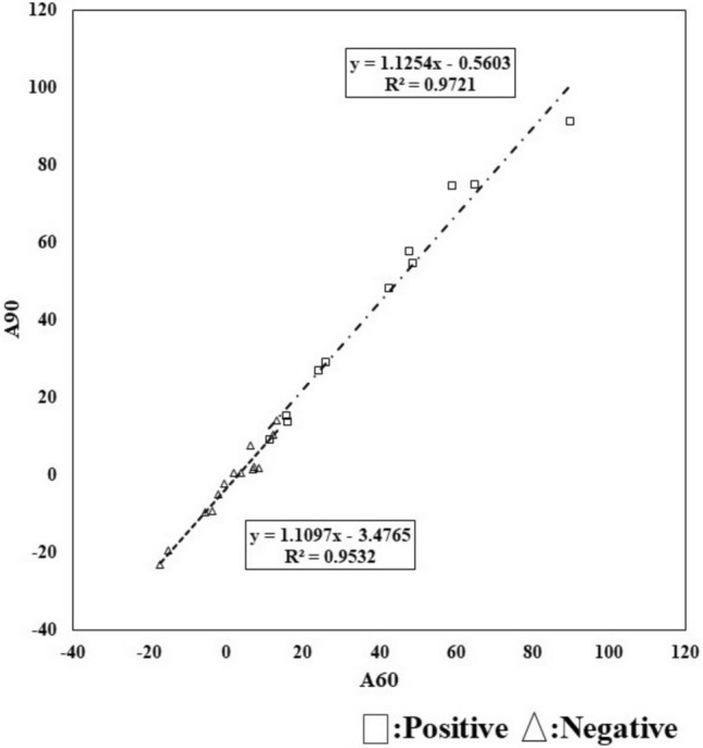
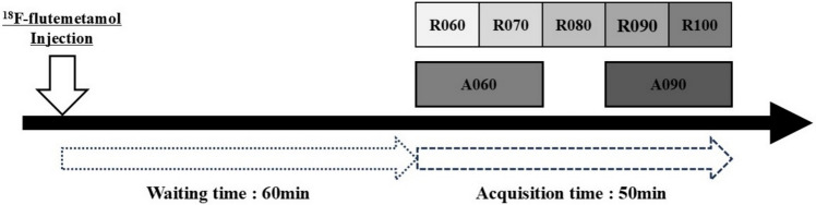
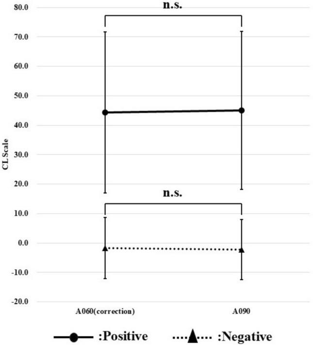
The study population consisted of 25 subjects who agreed to participate in the study. After visual evaluation by the radiologist, 14 subjects tested negative for Aβ accumulation, and 11 subjects tested positive as well. The study population was treated with 18F-flutemeamol, and list mode acquisition was performed for 50 min starting at 60 min after the time of administration. From the acquired list data, five PET images were extracted at 10-min intervals from the start to the end of acquisition, a PET image corresponding to 20 min of acquisition from 60 min after administration, and a PET image corresponding to 20 min of acquisition from 90 min after administration, respectively. Pixel values were measured for the PET images created and quantitative indices (pixel value, SUVr, Centiloid scale) were calculated and compared.
Results
In most of the PET images, pixel values showed a decreasing trend with elapsed time after radiopharmaceutical administration. Accordingly, calculated SUVr and Centiloid Scale also changed.
Conclusions
Elapsed time after radiopharmaceutical administration resulted in a washout of the radiopharmaceutical accumulation in the brain. From this, it was suggested that the quantitative indices change.
期刊介绍:
Annals of Nuclear Medicine is an official journal of the Japanese Society of Nuclear Medicine. It develops the appropriate application of radioactive substances and stable nuclides in the field of medicine.
The journal promotes the exchange of ideas and information and research in nuclear medicine and includes the medical application of radionuclides and related subjects. It presents original articles, short communications, reviews and letters to the editor.

 求助内容:
求助内容: 应助结果提醒方式:


