Gilbert M Schwarz, Alexander Synek, Stephanie Huber, Jochen G Hofstaetter, Dieter Pahr, Andreas Reisinger, Sylvia Nürnberger, Lena Hirtler
{"title":"头颅髓内钉去除后股骨骨折负荷降低:一项生物力学离体研究。","authors":"Gilbert M Schwarz, Alexander Synek, Stephanie Huber, Jochen G Hofstaetter, Dieter Pahr, Andreas Reisinger, Sylvia Nürnberger, Lena Hirtler","doi":"10.1302/2046-3758.145.BJR-2024-0278.R2","DOIUrl":null,"url":null,"abstract":"<p><strong>Aims: </strong>Spontaneous neck fractures are feared complications of cephalomedullary nail removal after successful healing of per- and subtrochanteric fractures. To date, the initial postoperative stability as well as the correct weightbearing regimen remain unclear. The aim of this biomechanical ex vivo study was to evaluate the initial postoperative failure load after hardware removal of specimens, which received cephalomedullary nails during their lifetime.</p><p><strong>Methods: </strong>A total of 20 specimens of voluntary body donors were included in this study. Group 1 (n = 10) consisted of specimens that received cephalomedullary nails during their lifetime due to per- or subtrochanteric fractures. Each individual was matched for age, sex, femur size, and neck-shaft angle (Group 2 = control, n = 10). Biomechanical testing was performed in a single-leg stance setting, and volumetric bone mineral density (vBMD) was measured proximally at the femoral neck and distally at the epicondyles.</p><p><strong>Results: </strong>Groups 1 and 2 differed significantly in terms of failure loads (p = 0.002), fracture types, and ratios of proximal and distal vBMD (p = 0.035). Femora after nail removal were significantly weaker (1,835.0 N vs 4,523.0 N) and showed lower ratios of proximal to distal vBMD (0.74 vs 1.18), which indicated altered stress distributions at the femoral neck in presence of femoral neck screws. They were further characterized by predominantly subcapital buckle-type fractures, while the control Group 2 showed predominantly transcervical fractures.</p><p><strong>Conclusion: </strong>Altered stress distribution in presence of femoral neck screws leads to changes in biomechanical properties of the proximal femur, resulting in potentially unstable situations after nail removal in clinical settings. Elective removal of cephalomedullary nails should be undertaken with caution in view of the potentially increased fracture risk.</p>","PeriodicalId":9074,"journal":{"name":"Bone & Joint Research","volume":"14 5","pages":"368-375"},"PeriodicalIF":5.1000,"publicationDate":"2025-05-01","publicationTypes":"Journal Article","fieldsOfStudy":null,"isOpenAccess":false,"openAccessPdf":"https://www.ncbi.nlm.nih.gov/pmc/articles/PMC12043369/pdf/","citationCount":"0","resultStr":"{\"title\":\"Decreased femoral fracture load after cephalomedullary nail removal : a biomechanical ex vivo study.\",\"authors\":\"Gilbert M Schwarz, Alexander Synek, Stephanie Huber, Jochen G Hofstaetter, Dieter Pahr, Andreas Reisinger, Sylvia Nürnberger, Lena Hirtler\",\"doi\":\"10.1302/2046-3758.145.BJR-2024-0278.R2\",\"DOIUrl\":null,\"url\":null,\"abstract\":\"<p><strong>Aims: </strong>Spontaneous neck fractures are feared complications of cephalomedullary nail removal after successful healing of per- and subtrochanteric fractures. To date, the initial postoperative stability as well as the correct weightbearing regimen remain unclear. The aim of this biomechanical ex vivo study was to evaluate the initial postoperative failure load after hardware removal of specimens, which received cephalomedullary nails during their lifetime.</p><p><strong>Methods: </strong>A total of 20 specimens of voluntary body donors were included in this study. Group 1 (n = 10) consisted of specimens that received cephalomedullary nails during their lifetime due to per- or subtrochanteric fractures. Each individual was matched for age, sex, femur size, and neck-shaft angle (Group 2 = control, n = 10). Biomechanical testing was performed in a single-leg stance setting, and volumetric bone mineral density (vBMD) was measured proximally at the femoral neck and distally at the epicondyles.</p><p><strong>Results: </strong>Groups 1 and 2 differed significantly in terms of failure loads (p = 0.002), fracture types, and ratios of proximal and distal vBMD (p = 0.035). Femora after nail removal were significantly weaker (1,835.0 N vs 4,523.0 N) and showed lower ratios of proximal to distal vBMD (0.74 vs 1.18), which indicated altered stress distributions at the femoral neck in presence of femoral neck screws. They were further characterized by predominantly subcapital buckle-type fractures, while the control Group 2 showed predominantly transcervical fractures.</p><p><strong>Conclusion: </strong>Altered stress distribution in presence of femoral neck screws leads to changes in biomechanical properties of the proximal femur, resulting in potentially unstable situations after nail removal in clinical settings. Elective removal of cephalomedullary nails should be undertaken with caution in view of the potentially increased fracture risk.</p>\",\"PeriodicalId\":9074,\"journal\":{\"name\":\"Bone & Joint Research\",\"volume\":\"14 5\",\"pages\":\"368-375\"},\"PeriodicalIF\":5.1000,\"publicationDate\":\"2025-05-01\",\"publicationTypes\":\"Journal Article\",\"fieldsOfStudy\":null,\"isOpenAccess\":false,\"openAccessPdf\":\"https://www.ncbi.nlm.nih.gov/pmc/articles/PMC12043369/pdf/\",\"citationCount\":\"0\",\"resultStr\":null,\"platform\":\"Semanticscholar\",\"paperid\":null,\"PeriodicalName\":\"Bone & Joint Research\",\"FirstCategoryId\":\"3\",\"ListUrlMain\":\"https://doi.org/10.1302/2046-3758.145.BJR-2024-0278.R2\",\"RegionNum\":2,\"RegionCategory\":\"医学\",\"ArticlePicture\":[],\"TitleCN\":null,\"AbstractTextCN\":null,\"PMCID\":null,\"EPubDate\":\"\",\"PubModel\":\"\",\"JCR\":\"Q2\",\"JCRName\":\"CELL & TISSUE ENGINEERING\",\"Score\":null,\"Total\":0}","platform":"Semanticscholar","paperid":null,"PeriodicalName":"Bone & Joint Research","FirstCategoryId":"3","ListUrlMain":"https://doi.org/10.1302/2046-3758.145.BJR-2024-0278.R2","RegionNum":2,"RegionCategory":"医学","ArticlePicture":[],"TitleCN":null,"AbstractTextCN":null,"PMCID":null,"EPubDate":"","PubModel":"","JCR":"Q2","JCRName":"CELL & TISSUE ENGINEERING","Score":null,"Total":0}
引用次数: 0
摘要
目的:股骨粗隆处及股骨粗隆下骨折成功愈合后,取下头髓钉后发生自发性颈骨折是常见的并发症。迄今为止,最初的术后稳定性以及正确的负重方案仍不清楚。这项生物力学离体研究的目的是评估在其一生中接受头髓钉的标本在硬体移除后的初始术后失效负荷。方法:本研究共纳入20例自愿遗体捐献者标本。第1组(n = 10)为因粗隆部或粗隆下骨折而在其一生中接受头髓内钉治疗的患者。每个个体根据年龄、性别、股骨大小和颈轴角度进行匹配(组2 =对照组,n = 10)。在单腿站立状态下进行生物力学测试,并在股骨颈近端和上髁远端测量体积骨密度(vBMD)。结果:1组和2组在骨折负荷(p = 0.002)、骨折类型、近端和远端vBMD比例(p = 0.035)方面差异有统计学意义。除钉后股骨明显较弱(1,835.0 N vs 4,523.0 N),近端与远端vBMD比例较低(0.74 vs 1.18),表明股骨颈螺钉存在时股骨颈应力分布发生了改变。他们进一步以亚资本屈曲型骨折为主,而对照组2则以经颈椎骨折为主。结论:股骨颈螺钉存在时应力分布的改变会导致股骨近端生物力学特性的改变,从而导致临床拔钉后潜在的不稳定情况。考虑到可能增加的骨折风险,应谨慎选择切除头髓钉。
Decreased femoral fracture load after cephalomedullary nail removal : a biomechanical ex vivo study.
Aims: Spontaneous neck fractures are feared complications of cephalomedullary nail removal after successful healing of per- and subtrochanteric fractures. To date, the initial postoperative stability as well as the correct weightbearing regimen remain unclear. The aim of this biomechanical ex vivo study was to evaluate the initial postoperative failure load after hardware removal of specimens, which received cephalomedullary nails during their lifetime.
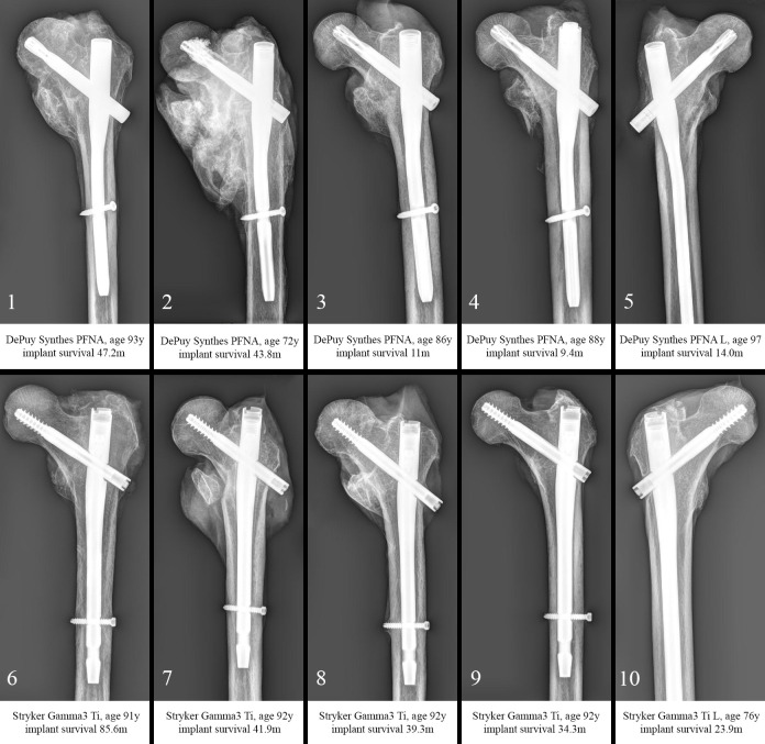
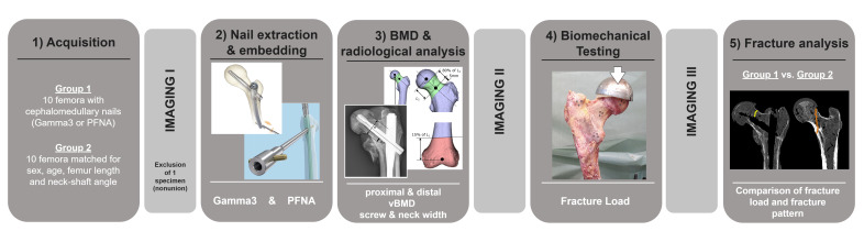
Methods: A total of 20 specimens of voluntary body donors were included in this study. Group 1 (n = 10) consisted of specimens that received cephalomedullary nails during their lifetime due to per- or subtrochanteric fractures. Each individual was matched for age, sex, femur size, and neck-shaft angle (Group 2 = control, n = 10). Biomechanical testing was performed in a single-leg stance setting, and volumetric bone mineral density (vBMD) was measured proximally at the femoral neck and distally at the epicondyles.
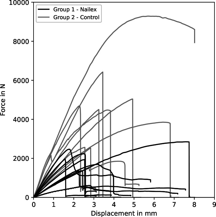
Results: Groups 1 and 2 differed significantly in terms of failure loads (p = 0.002), fracture types, and ratios of proximal and distal vBMD (p = 0.035). Femora after nail removal were significantly weaker (1,835.0 N vs 4,523.0 N) and showed lower ratios of proximal to distal vBMD (0.74 vs 1.18), which indicated altered stress distributions at the femoral neck in presence of femoral neck screws. They were further characterized by predominantly subcapital buckle-type fractures, while the control Group 2 showed predominantly transcervical fractures.
Conclusion: Altered stress distribution in presence of femoral neck screws leads to changes in biomechanical properties of the proximal femur, resulting in potentially unstable situations after nail removal in clinical settings. Elective removal of cephalomedullary nails should be undertaken with caution in view of the potentially increased fracture risk.
 求助内容:
求助内容: 应助结果提醒方式:


