Khizar Rana, David Curragh, Valerie Juniat, Sandy Patel, James Slattery, Dinesh Selva
{"title":"经蝶窦视神经管减压术治疗视神经管静脉畸形1例。","authors":"Khizar Rana, David Curragh, Valerie Juniat, Sandy Patel, James Slattery, Dinesh Selva","doi":"10.1159/000545542","DOIUrl":null,"url":null,"abstract":"<p><strong>Introduction: </strong>Intracanalicular vascular malformations are rare. This report describes a case of compressive optic neuropathy secondary to an intracanalicular venous malformation managed with endoscopic transsphenoidal optic canal decompression.</p><p><strong>Case presentation: </strong>A 43-year-old female presented with an 18-month history of painless vision loss secondary to an intracanalicular venous malformation causing compressive optic neuropathy. Ophthalmic examination showed reduced visual acuity and color vision, relative afferent pupillary defect, and optic disc pallor. Imaging findings were consistent with a slow-flow vascular malformation. An endoscopic transsphenoidal optic canal decompression was performed. The lesion was found to be wrapping around the optic nerve. At follow-up after 1 year, visual acuity had improved along with restoration of full color vision and visual fields.</p><p><strong>Conclusion: </strong>This case highlights the successful use of endoscopic transsphenoidal optic canal decompression to treat compressive optic neuropathy caused by an intracanalicular venous malformation.</p>","PeriodicalId":9635,"journal":{"name":"Case Reports in Ophthalmology","volume":"16 1","pages":"313-316"},"PeriodicalIF":0.6000,"publicationDate":"2025-04-03","publicationTypes":"Journal Article","fieldsOfStudy":null,"isOpenAccess":false,"openAccessPdf":"https://www.ncbi.nlm.nih.gov/pmc/articles/PMC12064154/pdf/","citationCount":"0","resultStr":"{\"title\":\"Transsphenoidal Optic Canal Decompression to Manage a Venous Malformation of the Optic Canal: A Case Report.\",\"authors\":\"Khizar Rana, David Curragh, Valerie Juniat, Sandy Patel, James Slattery, Dinesh Selva\",\"doi\":\"10.1159/000545542\",\"DOIUrl\":null,\"url\":null,\"abstract\":\"<p><strong>Introduction: </strong>Intracanalicular vascular malformations are rare. This report describes a case of compressive optic neuropathy secondary to an intracanalicular venous malformation managed with endoscopic transsphenoidal optic canal decompression.</p><p><strong>Case presentation: </strong>A 43-year-old female presented with an 18-month history of painless vision loss secondary to an intracanalicular venous malformation causing compressive optic neuropathy. Ophthalmic examination showed reduced visual acuity and color vision, relative afferent pupillary defect, and optic disc pallor. Imaging findings were consistent with a slow-flow vascular malformation. An endoscopic transsphenoidal optic canal decompression was performed. The lesion was found to be wrapping around the optic nerve. At follow-up after 1 year, visual acuity had improved along with restoration of full color vision and visual fields.</p><p><strong>Conclusion: </strong>This case highlights the successful use of endoscopic transsphenoidal optic canal decompression to treat compressive optic neuropathy caused by an intracanalicular venous malformation.</p>\",\"PeriodicalId\":9635,\"journal\":{\"name\":\"Case Reports in Ophthalmology\",\"volume\":\"16 1\",\"pages\":\"313-316\"},\"PeriodicalIF\":0.6000,\"publicationDate\":\"2025-04-03\",\"publicationTypes\":\"Journal Article\",\"fieldsOfStudy\":null,\"isOpenAccess\":false,\"openAccessPdf\":\"https://www.ncbi.nlm.nih.gov/pmc/articles/PMC12064154/pdf/\",\"citationCount\":\"0\",\"resultStr\":null,\"platform\":\"Semanticscholar\",\"paperid\":null,\"PeriodicalName\":\"Case Reports in Ophthalmology\",\"FirstCategoryId\":\"1085\",\"ListUrlMain\":\"https://doi.org/10.1159/000545542\",\"RegionNum\":0,\"RegionCategory\":null,\"ArticlePicture\":[],\"TitleCN\":null,\"AbstractTextCN\":null,\"PMCID\":null,\"EPubDate\":\"2025/1/1 0:00:00\",\"PubModel\":\"eCollection\",\"JCR\":\"Q4\",\"JCRName\":\"OPHTHALMOLOGY\",\"Score\":null,\"Total\":0}","platform":"Semanticscholar","paperid":null,"PeriodicalName":"Case Reports in Ophthalmology","FirstCategoryId":"1085","ListUrlMain":"https://doi.org/10.1159/000545542","RegionNum":0,"RegionCategory":null,"ArticlePicture":[],"TitleCN":null,"AbstractTextCN":null,"PMCID":null,"EPubDate":"2025/1/1 0:00:00","PubModel":"eCollection","JCR":"Q4","JCRName":"OPHTHALMOLOGY","Score":null,"Total":0}
Transsphenoidal Optic Canal Decompression to Manage a Venous Malformation of the Optic Canal: A Case Report.
Introduction: Intracanalicular vascular malformations are rare. This report describes a case of compressive optic neuropathy secondary to an intracanalicular venous malformation managed with endoscopic transsphenoidal optic canal decompression.
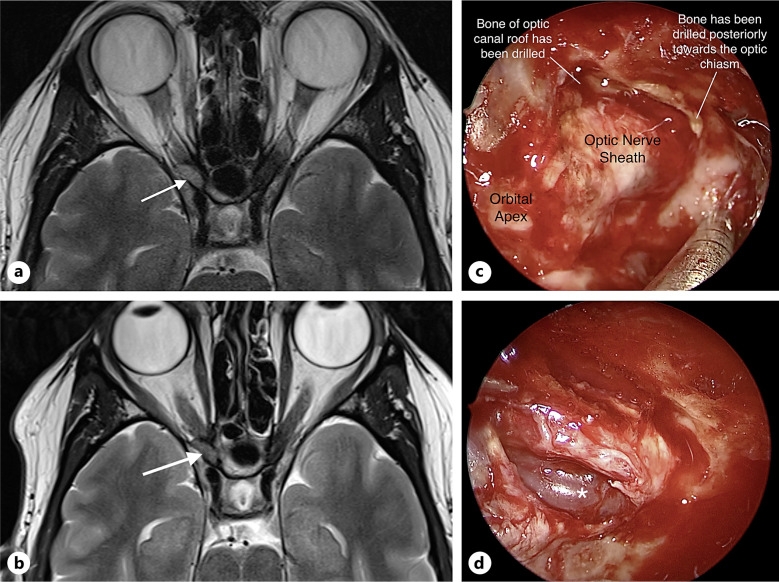
Case presentation: A 43-year-old female presented with an 18-month history of painless vision loss secondary to an intracanalicular venous malformation causing compressive optic neuropathy. Ophthalmic examination showed reduced visual acuity and color vision, relative afferent pupillary defect, and optic disc pallor. Imaging findings were consistent with a slow-flow vascular malformation. An endoscopic transsphenoidal optic canal decompression was performed. The lesion was found to be wrapping around the optic nerve. At follow-up after 1 year, visual acuity had improved along with restoration of full color vision and visual fields.
Conclusion: This case highlights the successful use of endoscopic transsphenoidal optic canal decompression to treat compressive optic neuropathy caused by an intracanalicular venous malformation.
期刊介绍:
This peer-reviewed online-only journal publishes original case reports covering the entire spectrum of ophthalmology, including prevention, diagnosis, treatment, toxicities of therapy, supportive care, quality-of-life, and survivorship issues. The submission of negative results is strongly encouraged. The journal will also accept case reports dealing with the use of novel technologies, both in the arena of diagnosis and treatment. Supplementary material is welcomed. The intent of the journal is to provide clinicians and researchers with a tool to disseminate their personal experiences to a wider public as well as to review interesting cases encountered by colleagues all over the world. Universally used terms can be searched across the entire growing collection of case reports, further facilitating the retrieval of specific information. Following the open access principle, the entire contents can be retrieved at no charge, guaranteeing easy access to this valuable source of anecdotal information at all times.

 求助内容:
求助内容: 应助结果提醒方式:


