Martha Fowler, Alvaro Moreno Lozano, Julian Krause, Patrick Bednarz, Shalini Pandey, Mina Ghayour, Qixu Zhang and Omid Veiseh
{"title":"通过构建的GelMA/PEGDA水凝胶引导血管浸润:通道直径、长度和复杂性的体内研究","authors":"Martha Fowler, Alvaro Moreno Lozano, Julian Krause, Patrick Bednarz, Shalini Pandey, Mina Ghayour, Qixu Zhang and Omid Veiseh","doi":"10.1039/D5BM00193E","DOIUrl":null,"url":null,"abstract":"<p >Organ shortages for transplantation in the United States impact over 100 000 patients, with 17 dying daily due to the lack of available organs. This growing need is exacerbated by the limited functionality and disease risk of donated organs. Tissue-engineered organs present a promising alternative, requiring optimized scaffold architecture and cell integration. Vascular networks within organs are essential for supplying oxygen and nutrients to cells, with a critical distance between blood vessels and surrounding tissue to allow effective diffusion. Various microfabrication techniques, such as electrospinning, freeze-drying, and gas foaming, have been employed to develop engineered organs. However, these techniques often lack the complexity needed to support vascularization. 3D bioprinting, particularly digital light projection (DLP)-based stereolithography, offers a solution by enabling high-resolution control of both external and internal architectures. Gelatin methacrylate (GelMA) and polyethylene glycol diacrylate (PEGDA) hydrogels have shown potential for tissue integration in simple structures but require further optimization for vascularization in more complex constructs. This study utilizes DLP to 3D bioprint GelMA/PEGDA hydrogels, exploring various channel designs to enhance tissue infiltration and vascularization in rodent models, providing a potential platform for cell and tissue transplantation. We demonstrate that GelMA/PEGDA hydrogels are mechanically robust, biocompatible, and support <em>in vivo</em> vascular infiltration. Channel diameter significantly influenced vascularization, with 1 mm channels yielding the highest infiltration, while channel length had minimal impact. Among five tested architectures, one design (GEO3) promoted the greatest vascular ingrowth, establishing a tunable hydrogel platform for prevascularized tissue engineering applications.</p>","PeriodicalId":65,"journal":{"name":"Biomaterials Science","volume":" 11","pages":" 2951-2960"},"PeriodicalIF":5.7000,"publicationDate":"2025-04-25","publicationTypes":"Journal Article","fieldsOfStudy":null,"isOpenAccess":false,"openAccessPdf":"https://pubs.rsc.org/en/content/articlepdf/2025/bm/d5bm00193e?page=search","citationCount":"0","resultStr":"{\"title\":\"Guiding vascular infiltration through architected GelMA/PEGDA hydrogels: an in vivo study of channel diameter, length, and complexity†\",\"authors\":\"Martha Fowler, Alvaro Moreno Lozano, Julian Krause, Patrick Bednarz, Shalini Pandey, Mina Ghayour, Qixu Zhang and Omid Veiseh\",\"doi\":\"10.1039/D5BM00193E\",\"DOIUrl\":null,\"url\":null,\"abstract\":\"<p >Organ shortages for transplantation in the United States impact over 100 000 patients, with 17 dying daily due to the lack of available organs. This growing need is exacerbated by the limited functionality and disease risk of donated organs. Tissue-engineered organs present a promising alternative, requiring optimized scaffold architecture and cell integration. Vascular networks within organs are essential for supplying oxygen and nutrients to cells, with a critical distance between blood vessels and surrounding tissue to allow effective diffusion. Various microfabrication techniques, such as electrospinning, freeze-drying, and gas foaming, have been employed to develop engineered organs. However, these techniques often lack the complexity needed to support vascularization. 3D bioprinting, particularly digital light projection (DLP)-based stereolithography, offers a solution by enabling high-resolution control of both external and internal architectures. Gelatin methacrylate (GelMA) and polyethylene glycol diacrylate (PEGDA) hydrogels have shown potential for tissue integration in simple structures but require further optimization for vascularization in more complex constructs. This study utilizes DLP to 3D bioprint GelMA/PEGDA hydrogels, exploring various channel designs to enhance tissue infiltration and vascularization in rodent models, providing a potential platform for cell and tissue transplantation. We demonstrate that GelMA/PEGDA hydrogels are mechanically robust, biocompatible, and support <em>in vivo</em> vascular infiltration. Channel diameter significantly influenced vascularization, with 1 mm channels yielding the highest infiltration, while channel length had minimal impact. Among five tested architectures, one design (GEO3) promoted the greatest vascular ingrowth, establishing a tunable hydrogel platform for prevascularized tissue engineering applications.</p>\",\"PeriodicalId\":65,\"journal\":{\"name\":\"Biomaterials Science\",\"volume\":\" 11\",\"pages\":\" 2951-2960\"},\"PeriodicalIF\":5.7000,\"publicationDate\":\"2025-04-25\",\"publicationTypes\":\"Journal Article\",\"fieldsOfStudy\":null,\"isOpenAccess\":false,\"openAccessPdf\":\"https://pubs.rsc.org/en/content/articlepdf/2025/bm/d5bm00193e?page=search\",\"citationCount\":\"0\",\"resultStr\":null,\"platform\":\"Semanticscholar\",\"paperid\":null,\"PeriodicalName\":\"Biomaterials Science\",\"FirstCategoryId\":\"5\",\"ListUrlMain\":\"https://pubs.rsc.org/en/content/articlelanding/2025/bm/d5bm00193e\",\"RegionNum\":3,\"RegionCategory\":\"医学\",\"ArticlePicture\":[],\"TitleCN\":null,\"AbstractTextCN\":null,\"PMCID\":null,\"EPubDate\":\"\",\"PubModel\":\"\",\"JCR\":\"Q1\",\"JCRName\":\"MATERIALS SCIENCE, BIOMATERIALS\",\"Score\":null,\"Total\":0}","platform":"Semanticscholar","paperid":null,"PeriodicalName":"Biomaterials Science","FirstCategoryId":"5","ListUrlMain":"https://pubs.rsc.org/en/content/articlelanding/2025/bm/d5bm00193e","RegionNum":3,"RegionCategory":"医学","ArticlePicture":[],"TitleCN":null,"AbstractTextCN":null,"PMCID":null,"EPubDate":"","PubModel":"","JCR":"Q1","JCRName":"MATERIALS SCIENCE, BIOMATERIALS","Score":null,"Total":0}
Guiding vascular infiltration through architected GelMA/PEGDA hydrogels: an in vivo study of channel diameter, length, and complexity†
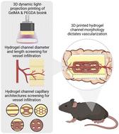
Organ shortages for transplantation in the United States impact over 100 000 patients, with 17 dying daily due to the lack of available organs. This growing need is exacerbated by the limited functionality and disease risk of donated organs. Tissue-engineered organs present a promising alternative, requiring optimized scaffold architecture and cell integration. Vascular networks within organs are essential for supplying oxygen and nutrients to cells, with a critical distance between blood vessels and surrounding tissue to allow effective diffusion. Various microfabrication techniques, such as electrospinning, freeze-drying, and gas foaming, have been employed to develop engineered organs. However, these techniques often lack the complexity needed to support vascularization. 3D bioprinting, particularly digital light projection (DLP)-based stereolithography, offers a solution by enabling high-resolution control of both external and internal architectures. Gelatin methacrylate (GelMA) and polyethylene glycol diacrylate (PEGDA) hydrogels have shown potential for tissue integration in simple structures but require further optimization for vascularization in more complex constructs. This study utilizes DLP to 3D bioprint GelMA/PEGDA hydrogels, exploring various channel designs to enhance tissue infiltration and vascularization in rodent models, providing a potential platform for cell and tissue transplantation. We demonstrate that GelMA/PEGDA hydrogels are mechanically robust, biocompatible, and support in vivo vascular infiltration. Channel diameter significantly influenced vascularization, with 1 mm channels yielding the highest infiltration, while channel length had minimal impact. Among five tested architectures, one design (GEO3) promoted the greatest vascular ingrowth, establishing a tunable hydrogel platform for prevascularized tissue engineering applications.
期刊介绍:
Biomaterials Science is an international high impact journal exploring the science of biomaterials and their translation towards clinical use. Its scope encompasses new concepts in biomaterials design, studies into the interaction of biomaterials with the body, and the use of materials to answer fundamental biological questions.
 求助内容:
求助内容: 应助结果提醒方式:


